Cyst, Mass, Nodule, Lesion: What the Words Mean
Radiology reports use cyst, mass, nodule, and lesion as descriptive labels — not interchangeable diagnoses. Patient-facing report guides and thoracic imaging glossaries explain how these words are typically used.
Lesion is a broad umbrella word for an abnormal-appearing area. Cyst usually describes a fluid-filled (or air-filled, in some lung contexts) structure with a defined wall. Nodule and mass are size-based descriptors used especially in chest imaging: in Fleischner Society thoracic terminology, a nodule is typically ≤30 mm and a mass is >30 mm. Exact meanings can vary by body part and modality. Only your clinician can explain what a word means on your study.
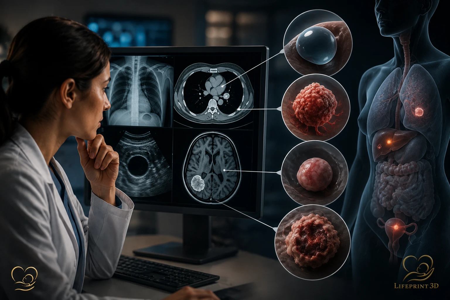
Seeing cyst, mass, nodule, or lesion on a radiology report can feel alarming — especially when those words appear online next to worst-case stories. In imaging language, these terms are usually descriptive labels for how something looks on the study, not a finished diagnosis of cancer or another specific disease. This article explains how those words are commonly used in educational and professional glossaries. It cannot interpret your personal report.
These Words Describe Appearance — Not Your Diagnosis
RadiologyInfo.org explains that radiology reports are written primarily for the ordering clinician and often use specialized terms that can be confusing when patients read them first in a portal. The report’s Impression summarizes important findings; wording there still needs clinical context.
Cyst, mass, nodule, and lesion are imaging descriptors. They do not automatically mean cancer, infection, or “nothing to worry about.” Severity and next steps depend on location, size, borders, enhancement, comparison with prior studies, and your history — judgments that belong with your care team.
Lesion: The Broadest Label
Lesion is an umbrella term for an abnormal-appearing area. Radiologists may use it when they want a neutral word before more specific descriptors are available, or when several possibilities remain open.
If a report says “lesion,” look at the rest of the sentence: size, organ, simple vs complex, and any recommended follow-up. The word alone does not rank urgency.
Cyst: Usually Fluid-Filled (With Important Exceptions)
In everyday abdominal and pelvic imaging education, cyst often means a roughly round structure that behaves like fluid on ultrasound, CT, or MRI — for example many simple kidney or liver cysts discussed as common incidental findings in RadiologyInfo report examples.
In thoracic (chest) imaging, the Fleischner Society glossary defines cyst in a more lung-specific way (including air-containing structures in lung parenchyma). That is a reminder that the same English word can carry different technical definitions by body region. Do not paste a lung glossary definition onto a kidney finding — ask your clinician.
Nodule and Mass: Often About Size and Shape
The 2024 Fleischner Society Glossary of Terms for Thoracic Imaging defines a pulmonary nodule as a circumscribed, typically round opacity ≤30 mm in average diameter, and a mass as a circumscribed lesion greater than 30 mm. A rounded lesion larger than 30 mm is called a mass rather than a nodule in that framework.
Those size cutoffs are standardized for chest CT communication. Other organs may use nodule more loosely for a small rounded focus. Mass can also appear outside the chest as a descriptive word for a space-occupying finding — again without automatically meaning malignancy.
How to Read the Rest of the Sentence
Helpful companions to these nouns include adjectives and follow-up language: simple, complex, well-circumscribed, irregular, enhancing, stable compared with prior, too small to characterize, or recommend clinical correlation. RadiologyInfo notes that many findings are incidental and may cause no harm — and that radiologists may recommend correlating imaging with symptoms or labs.
For related education on when imaging may not be the next step, see why a clinician might recommend no imaging, and for spine-related wording that often worries patients, see degenerative changes on imaging.
When a Physical Model Can Aid Understanding
Report words such as cyst, nodule, mass, or lesion describe appearance on images — they are not themselves a 3D object you can hold. When a clinician already has volumetric CT or MRI of the region discussed, a digital 3D reconstruction or a physical printed anatomical model made from that existing dataset can sometimes help a care team or patient visualize spatial relationships that are hard to picture from a flat report.
A model is an educational or communication aid. It is not a diagnostic substitute for the radiology report, and it does not turn a descriptive word into a disease name. Services that print from imaging you already have should never be a reason to request a new scan.
References
RadiologyInfo.org (ACR/RSNA) — All About Your Radiology Report: What to Know (https://www.radiologyinfo.org/en/info/all-about-your-radiology-report).
RadiologyInfo.org — How to discuss radiology report results with your doctor (https://www.radiologyinfo.org/en/info/article-discussing-report-results).
Bankier AA, et al. — Fleischner Society: Glossary of Terms for Thoracic Imaging. Radiology. 2024 (PMID 38411514; doi: 10.1148/radiol.232558).
Hansell DM, et al. — Fleischner Society: glossary of terms for thoracic imaging. Radiology. 2008 (PMID 18195376; doi: 10.1148/radiol.2462070712).
Key Takeaways
- RadiologyInfo.
- Lesion is an umbrella term for an abnormal-appearing area.
- In everyday abdominal and pelvic imaging education, cyst often means a roughly round structure that behaves like fluid on ultrasound, CT, or MRI — for example many simple kidney or liver cysts discussed as common incidental findings in RadiologyInfo report examples.
- The 2024 Fleischner Society Glossary of Terms for Thoracic Imaging defines a pulmonary nodule as a circumscribed, typically round opacity ≤30 mm in average diameter, and a mass as a circumscribed lesion greater than 30 mm.
- Helpful companions to these nouns include adjectives and follow-up language: simple, complex, well-circumscribed, irregular, enhancing, stable compared with prior, too small to characterize, or recommend clinical correlation.
Frequently Asked Questions
Does “mass” mean cancer?
No. In imaging language, mass is often a size or morphology descriptor. Cancer is a pathologic or clinical diagnosis that requires more than a single word on a report.
Is a nodule always something that needs surgery?
No. Many nodules are monitored or further characterized based on guidelines and clinical context. This page cannot say what your nodule needs.
Why do definitions differ between lung and other organs?
Professional societies publish glossaries for specific imaging domains (such as thoracic CT). The same English word can be defined differently by body region.
Related articles
Glossary · Frequently asked questions · All Learning Center articles