Why Does My Baby Look Different on Ultrasound?
Ultrasound images are not photographs. Learn why your baby's face can look different between 2D, 3D and 4D scans and why appearance changes with gestational age.
Your baby can look different from one ultrasound image to another because ultrasound is a reconstruction of sound-wave data, not a photograph. The displayed appearance depends on imaging plane, fetal position, gestational age, surrounding structures, machine settings and, for 3D images, the rendering process. [11, 12, 13, 14]
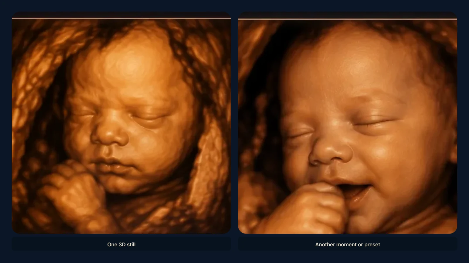
Ultrasound images are not photographs. Learn why your baby's face can look different between 2D, 3D and 4D scans and why appearance changes with gestational age.
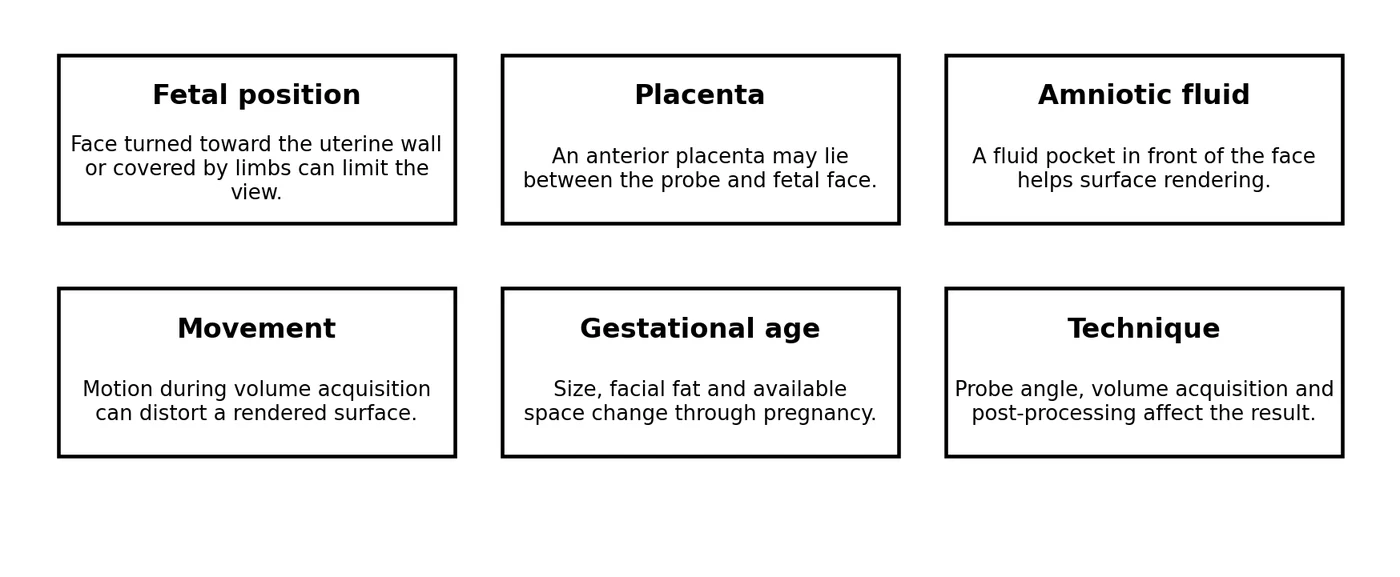
A 2D slice can change the apparent shape
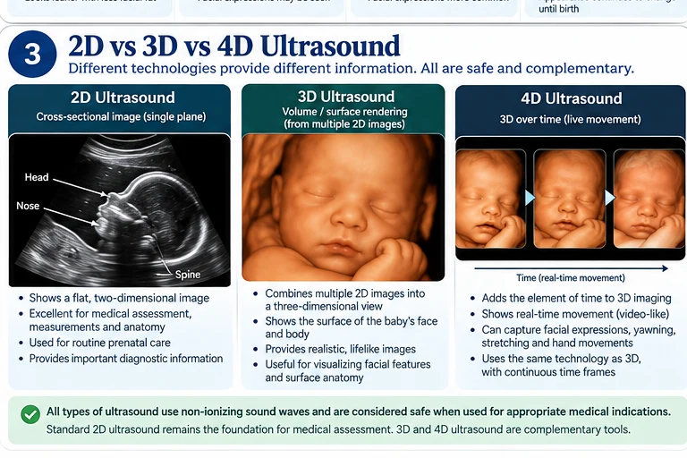
Two-dimensional ultrasound displays a thin cross-sectional plane. A slightly different angle through the nose, lips or forehead can make proportions look different. This is similar to slicing through a three-dimensional object at different angles: each slice is real, but no single slice represents the whole surface.
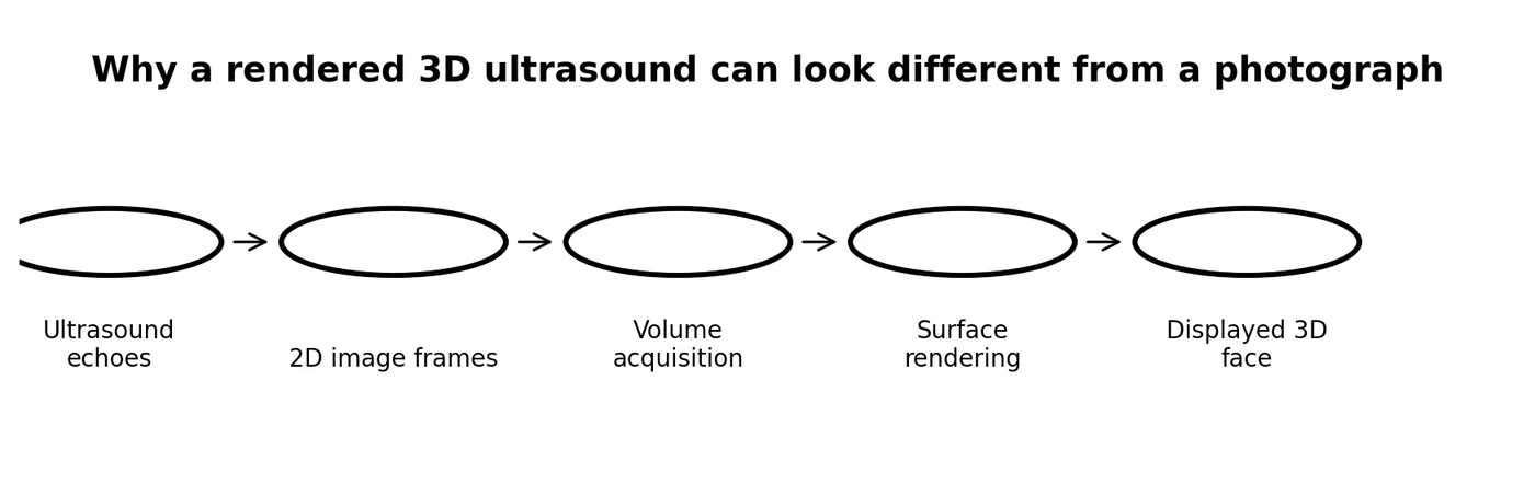
3D rendering adds another processing step
Three-dimensional ultrasound combines a volume of data and then displays selected surfaces. The software must decide which echoes represent the visible surface. Tissue touching the face can be included, while missing or shadowed data can be excluded. The result may look surprisingly realistic, but it remains a rendered medical image.
Gestational age changes the face
The fetal face grows throughout pregnancy. Later in gestation, increasing soft tissue and fat can produce fuller cheeks and a rounder contour. Research using 3D facial landmarks has documented measurable changes across gestation. [9, 17]
Expressions and motion can change a single frame
4D studies demonstrate fetal mouthing, yawning and other facial movements. A single frame captured during movement may therefore look very different from another frame seconds later. These movements should not be interpreted as adult-like emotions. [16]
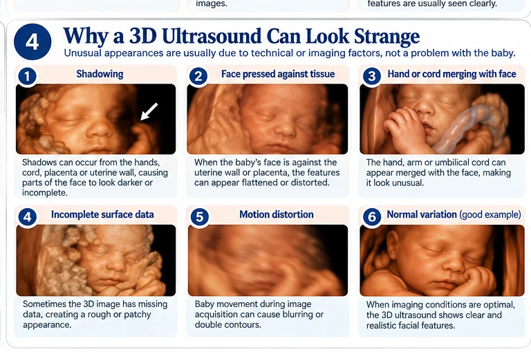
Why ultrasound and the newborn may not look identical
The intrauterine face is influenced by gestational age, position and surrounding tissues. The ultrasound rendering is also limited by resolution and acquisition geometry. After birth, lighting, gravity, facial movement and continued maturation change appearance further. Ultrasound should therefore be viewed as an imaging representation, not a precise facial forecast.
Why 3D physical visualization can still be meaningful
Although a rendering is not a photograph, a well-acquired 3D dataset contains spatial information. That can be valuable for understanding surface form. A carefully produced physical representation can translate those captured spatial relationships into something tangible, provided the model does not imply detail absent from the source data.
Medical disclaimer
This article is intended for educational purposes only and does not provide medical advice, diagnosis, or treatment. Always consult a qualified healthcare professional regarding your individual medical care. This content is based on peer-reviewed scientific literature and current clinical guidelines available at the time of publication. Every article requires physician review before publication.
Key Takeaways
- Two-dimensional ultrasound displays a thin cross-sectional plane.
- Three-dimensional ultrasound combines a volume of data and then displays selected surfaces.
- The fetal face grows throughout pregnancy.
- 4D studies demonstrate fetal mouthing, yawning and other facial movements.
- The intrauterine face is influenced by gestational age, position and surrounding tissues.
Frequently Asked Questions
Why does my baby look different between scans?
Different imaging planes, gestational age, position, motion and rendering can all change appearance.
Why does 3D look more realistic than 2D?
3D can display an external surface, while 2D shows cross-sectional grayscale planes.
Will the newborn look exactly like the 3D scan?
No. The scan is a limited rendered representation and postnatal appearance also changes with lighting, movement and maturation.
References
- ACOG. Ultrasound Exams. American College of Obstetricians and Gynecologists. Standard obstetric ultrasound is commonly performed at 18-22 weeks; specialized techniques may include 3D ultrasound. https://www.acog.org/womens-health/faqs/ultrasound-exams
- ISUOG Practice Guidelines (updated): performance of the routine mid-trimester fetal ultrasound scan. Ultrasound Obstet Gynecol. 2022;59:840-856. https://www.isuog.org/resource/isuog-practice-guidelines-updated-performance-of-the-routine-mid-trimester-fetal-ultrasound-scan.html
- FDA. Ultrasound Imaging. U.S. Food and Drug Administration. Ultrasound does not use ionizing radiation; keepsake images are reasonable when produced during a medically indicated examination without additional exposure. https://www.fda.gov/radiation-emitting-products/medical-imaging/ultrasound-imaging
- AIUM. Prudent Clinical Use and Safety of Diagnostic Ultrasound. 2019. Supports prudent, medically appropriate use and attention to acoustic output and exposure time. https://www.aium.org/resources/official-statements/view/prudent-clinical-use-and-safety-of-diagnostic-ultrasound
- AIUM. Prudent Use and Safety of Diagnostic Ultrasound in Pregnancy. Official statement. https://www.aium.org/resources/official-statements/obstetric-ultrasound
- NHS. 20-week screening scan. The anomaly scan is usually performed between 18 and 21 weeks and assesses fetal anatomy and growth. https://www.nhs.uk/pregnancy/your-pregnancy-care/20-week-scan/
- MedlinePlus. Fetal development. U.S. National Library of Medicine. https://medlineplus.gov/ency/article/002398.htm
- Connor A, Matthew J, Chung E, Wright R, Rutherford MA. Factors influencing the successful acquisition and quality of three-dimensional souvenir obstetric ultrasound facial images: a narrative review and quantitative evaluation. Ultrasound. 2026. PMID: 42294041. DOI: 10.1177/1742271X261436619. DOI: 10.1177/1742271X261436619 PMID: 42294041 Source
- Accuracy and repeatability of fetal facial measurements in 3D ultrasound: A longitudinal study. Early Hum Dev. 2024. PMID: 38701668. DOI: 10.1016/j.earlhumdev.2024.106021. DOI: 10.1016/j.earlhumdev.2024.106021 PMID: 38701668 Source
- Three-dimensional sonographic visualization of the fetal face. AJR Am J Roentgenol. PMID: 9456969. DOI: 10.2214/ajr.170.2.9456969. DOI: 10.2214/ajr.170.2.9456969 PMID: 9456969 Source
- Two- and three-dimensional sonographic assessment of the fetal face. 1. A systematic analysis of the normal face. Ultrasound Obstet Gynecol. PMID: 15027008. DOI: 10.1002/uog.984. DOI: 10.1002/uog.984 PMID: 15027008 Source
- Diagnostic evaluation of the fetal face using 3-dimensional ultrasound. Ultrasound Q. 2008;24:215-223. PMID: 19060688. DOI: 10.1097/RUQ.0b013e31819073c2. DOI: 10.1097/RUQ.0b013e31819073c2 PMID: 19060688 Source
- Three-dimensional and four-dimensional ultrasound applications in fetal medicine. Curr Opin Obstet Gynecol. 2009;21:167-174. PMID: 19996869. DOI: 10.1097/GCO.0b013e328329243c. DOI: 10.1097/GCO.0b013e328329243c PMID: 19996869 Source
- Role of 3-D ultrasound in clinical obstetric practice: evolution over 20 years. Ultrasound Med Biol. 2015. PMID: 25748522. DOI: 10.1016/j.ultrasmedbio.2014.12.009. DOI: 10.1016/j.ultrasmedbio.2014.12.009 PMID: 25748522 Source
- 3D/4D sonography - any safety problem. J Perinat Med. 2016;44:125-129. PMID: 26376219. DOI: 10.1515/jpm-2015-0225. DOI: 10.1515/jpm-2015-0225 PMID: 26376219 Source
- 4D ultrasound study of fetal facial expressions at 20-24 weeks of gestation. Int J Gynaecol Obstet. 2014;126:275-279. PMID: 24996686. DOI: 10.1016/j.ijgo.2014.03.036. DOI: 10.1016/j.ijgo.2014.03.036 PMID: 24996686 Source
- Fetal face shape analysis from prenatal 3D ultrasound images. Sci Rep. 2024. PMID: 38388522. PMCID: PMC10884000. DOI: 10.1038/s41598-023-50386-9. DOI: 10.1038/s41598-023-50386-9 PMID: 38388522 Source
- Three-dimensional ultrasonic images of normal fetus. Study of pregnancies 20-36 weeks; satisfactory recognizable images were reported particularly from 24-34 weeks. PMID page. PMID: 11601189 Source
- Three-dimensional ultrasonographic assessments of fetal development. PMID: 9469279. PMID: 9469279 Source
- Three-dimensional reconstruction of fetal abnormalities using ultrasonography and magnetic resonance imaging. PMID: 29656679. PMID: 29656679 Source
Related articles
Glossary · Frequently asked questions · All Learning Center articles