Why Is My Baby's Face Hard to See on Ultrasound?
A baby's face can be difficult to see on ultrasound because of position, placenta, hands, fluid, movement or imaging geometry. Learn what these factors mean.
A difficult facial view is common and usually reflects imaging conditions rather than a problem with the baby. Ultrasound needs a useful acoustic path and, for 3D surface rendering, a separable surface in front of the face. Fetal position, placenta, limbs, umbilical cord, fluid, movement and technical settings can all interfere. [8, 20]
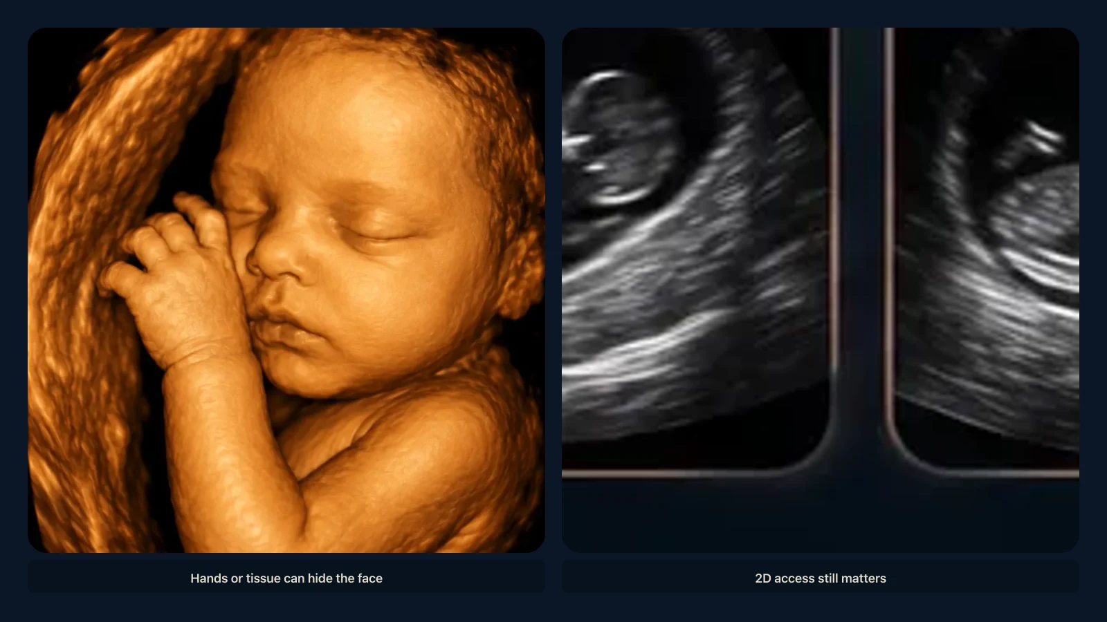
A baby's face can be difficult to see on ultrasound because of position, placenta, hands, fluid, movement or imaging geometry. Learn what these factors mean.
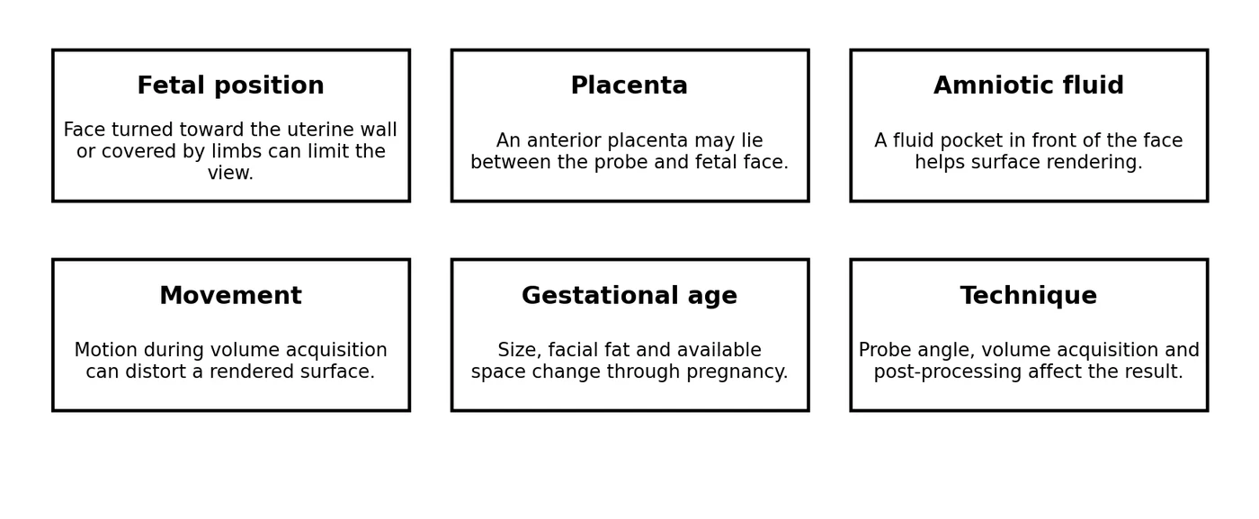
Fetal position
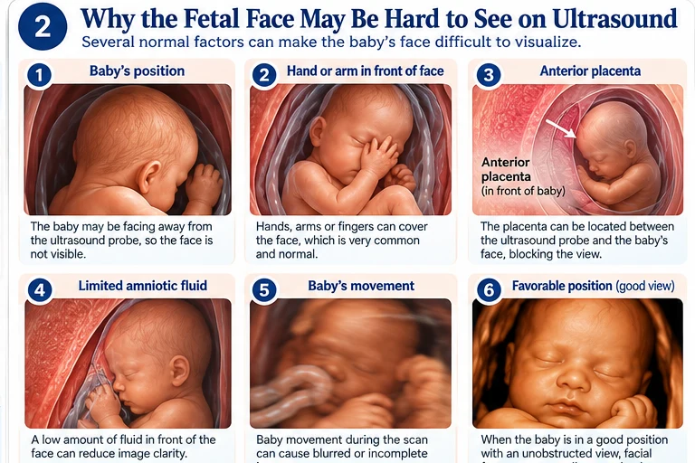
If the fetus is facing the maternal spine or uterine wall, the transducer may not have a direct view of the facial surface. A side-facing fetus may provide a good profile but a poor frontal rendering. Position can change during the examination or between visits.
Hands and feet near the face
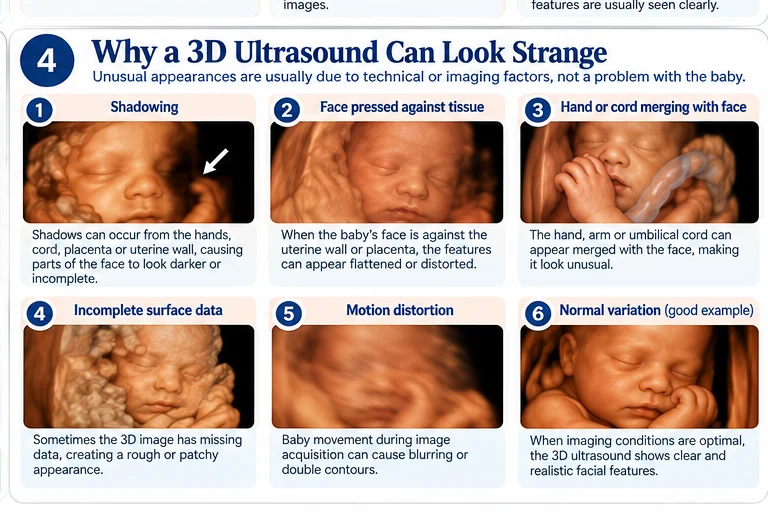
Fetuses commonly bring their hands and arms toward the face. On a 3D rendering, adjacent fingers or forearms may merge visually with the cheeks, mouth or forehead because the software is displaying surfaces that lie close together.
Placenta in front of the face
An anterior placenta is located on the front wall of the uterus. Depending on fetal position, placental tissue may lie between the transducer and face or directly against the facial surface. Recent quantitative work found placental site associated with 3D facial image quality. [8]
Amniotic fluid and surface rendering
A visible fluid space in front of the face helps the software distinguish the fetal surface from surrounding tissue. When that interface is narrow, parts of the nose, lips or chin can look incomplete. This is an imaging limitation, not a reason to diagnose low amniotic fluid from a souvenir image.
Movement and acquisition
A 3D volume is acquired over a short interval. If the fetus moves during acquisition, different portions of the volume may not align perfectly, creating distortion. Four-dimensional imaging repeats these acquisitions, but motion and obstruction can still limit quality. [15]
What matters clinically
A poor 3D portrait does not mean the diagnostic examination was poor. Clinicians can use multiple 2D planes and other techniques to assess fetal anatomy. If a required diagnostic structure cannot be adequately evaluated, the clinical team decides whether additional imaging is appropriate. [1, 2]
Medical disclaimer
This article is intended for educational purposes only and does not provide medical advice, diagnosis, or treatment. Always consult a qualified healthcare professional regarding your individual medical care. This content is based on peer-reviewed scientific literature and current clinical guidelines available at the time of publication. Every article requires physician review before publication.
Key Takeaways
- If the fetus is facing the maternal spine or uterine wall, the transducer may not have a direct view of the facial surface.
- Fetuses commonly bring their hands and arms toward the face.
- An anterior placenta is located on the front wall of the uterus.
- A visible fluid space in front of the face helps the software distinguish the fetal surface from surrounding tissue.
- A 3D volume is acquired over a short interval.
Frequently Asked Questions
Does a hidden face mean something is wrong?
Usually it reflects fetal position or imaging conditions. Clinical concerns are determined from the full examination.
Can the placenta block the face?
It can lie in front of or against the face depending on placental and fetal position.
Can a hand make the face look distorted?
Yes. Nearby limbs can merge with the facial surface on 3D rendering.
References
- ACOG. Ultrasound Exams. American College of Obstetricians and Gynecologists. Standard obstetric ultrasound is commonly performed at 18-22 weeks; specialized techniques may include 3D ultrasound. https://www.acog.org/womens-health/faqs/ultrasound-exams
- ISUOG Practice Guidelines (updated): performance of the routine mid-trimester fetal ultrasound scan. Ultrasound Obstet Gynecol. 2022;59:840-856. https://www.isuog.org/resource/isuog-practice-guidelines-updated-performance-of-the-routine-mid-trimester-fetal-ultrasound-scan.html
- FDA. Ultrasound Imaging. U.S. Food and Drug Administration. Ultrasound does not use ionizing radiation; keepsake images are reasonable when produced during a medically indicated examination without additional exposure. https://www.fda.gov/radiation-emitting-products/medical-imaging/ultrasound-imaging
- AIUM. Prudent Clinical Use and Safety of Diagnostic Ultrasound. 2019. Supports prudent, medically appropriate use and attention to acoustic output and exposure time. https://www.aium.org/resources/official-statements/view/prudent-clinical-use-and-safety-of-diagnostic-ultrasound
- AIUM. Prudent Use and Safety of Diagnostic Ultrasound in Pregnancy. Official statement. https://www.aium.org/resources/official-statements/obstetric-ultrasound
- NHS. 20-week screening scan. The anomaly scan is usually performed between 18 and 21 weeks and assesses fetal anatomy and growth. https://www.nhs.uk/pregnancy/your-pregnancy-care/20-week-scan/
- MedlinePlus. Fetal development. U.S. National Library of Medicine. https://medlineplus.gov/ency/article/002398.htm
- Connor A, Matthew J, Chung E, Wright R, Rutherford MA. Factors influencing the successful acquisition and quality of three-dimensional souvenir obstetric ultrasound facial images: a narrative review and quantitative evaluation. Ultrasound. 2026. PMID: 42294041. DOI: 10.1177/1742271X261436619. DOI: 10.1177/1742271X261436619 PMID: 42294041 Source
- Accuracy and repeatability of fetal facial measurements in 3D ultrasound: A longitudinal study. Early Hum Dev. 2024. PMID: 38701668. DOI: 10.1016/j.earlhumdev.2024.106021. DOI: 10.1016/j.earlhumdev.2024.106021 PMID: 38701668 Source
- Three-dimensional sonographic visualization of the fetal face. AJR Am J Roentgenol. PMID: 9456969. DOI: 10.2214/ajr.170.2.9456969. DOI: 10.2214/ajr.170.2.9456969 PMID: 9456969 Source
- Two- and three-dimensional sonographic assessment of the fetal face. 1. A systematic analysis of the normal face. Ultrasound Obstet Gynecol. PMID: 15027008. DOI: 10.1002/uog.984. DOI: 10.1002/uog.984 PMID: 15027008 Source
- Diagnostic evaluation of the fetal face using 3-dimensional ultrasound. Ultrasound Q. 2008;24:215-223. PMID: 19060688. DOI: 10.1097/RUQ.0b013e31819073c2. DOI: 10.1097/RUQ.0b013e31819073c2 PMID: 19060688 Source
- Three-dimensional and four-dimensional ultrasound applications in fetal medicine. Curr Opin Obstet Gynecol. 2009;21:167-174. PMID: 19996869. DOI: 10.1097/GCO.0b013e328329243c. DOI: 10.1097/GCO.0b013e328329243c PMID: 19996869 Source
- Role of 3-D ultrasound in clinical obstetric practice: evolution over 20 years. Ultrasound Med Biol. 2015. PMID: 25748522. DOI: 10.1016/j.ultrasmedbio.2014.12.009. DOI: 10.1016/j.ultrasmedbio.2014.12.009 PMID: 25748522 Source
- 3D/4D sonography - any safety problem. J Perinat Med. 2016;44:125-129. PMID: 26376219. DOI: 10.1515/jpm-2015-0225. DOI: 10.1515/jpm-2015-0225 PMID: 26376219 Source
- 4D ultrasound study of fetal facial expressions at 20-24 weeks of gestation. Int J Gynaecol Obstet. 2014;126:275-279. PMID: 24996686. DOI: 10.1016/j.ijgo.2014.03.036. DOI: 10.1016/j.ijgo.2014.03.036 PMID: 24996686 Source
- Fetal face shape analysis from prenatal 3D ultrasound images. Sci Rep. 2024. PMID: 38388522. PMCID: PMC10884000. DOI: 10.1038/s41598-023-50386-9. DOI: 10.1038/s41598-023-50386-9 PMID: 38388522 Source
- Three-dimensional ultrasonic images of normal fetus. Study of pregnancies 20-36 weeks; satisfactory recognizable images were reported particularly from 24-34 weeks. PMID page. PMID: 11601189 Source
- Three-dimensional ultrasonographic assessments of fetal development. PMID: 9469279. PMID: 9469279 Source
- Three-dimensional reconstruction of fetal abnormalities using ultrasonography and magnetic resonance imaging. PMID: 29656679. PMID: 29656679 Source
Related articles
Glossary · Frequently asked questions · All Learning Center articles