How Gallstones Are Found
Ultrasound is the most common first imaging test used to look for gallstones. RadiologyInfo and ACR appropriateness summaries explain how ultrasound, CT, MRCP, and related tests enter the pathway — without covering treatment.
Abdominal ultrasound is the most common test used to evaluate gallbladder abnormalities and is often the first imaging test for suspected gallstones or cholecystitis. RadiologyInfo also describes CT and MRCP as additional options clinicians may use. ACR Appropriateness Criteria materials on right-upper-quadrant pain similarly rate ultrasound as usually appropriate first-line imaging in common biliary scenarios. Your clinician chooses the pathway for your symptoms.
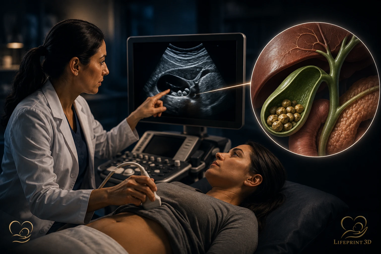
Gallstones are often discovered when someone has right-upper abdominal symptoms — and sometimes as an incidental finding on imaging done for another reason. This article explains how clinicians typically look for gallstones with imaging, based on RadiologyInfo patient education and ACR appropriateness materials. It covers detection, not diet, medications, ERCP technique details as therapy, or whether you personally need gallbladder surgery.
Imaging Education Only — Not Treatment Advice
We explain how stones and related gallbladder findings are looked for on imaging. We do not advise on pain control, antibiotics, dietary fat restriction, or surgical removal of the gallbladder.
Severe abdominal pain, fever, jaundice, or vomiting that worries you needs urgent clinical care — not triage from an educational webpage.
Why Ultrasound Is Usually First
RadiologyInfo’s gallstones page states that abdominal ultrasound produces pictures of the gallbladder and bile ducts, can show signs of inflammation or bile-flow blockage, and is the most common test performed to evaluate gallbladder abnormalities.
RadiologyInfo’s cholecystitis page similarly notes that ultrasound is often the first test done to evaluate for cholecystitis, uses sound waves without ionizing radiation, and is very good at showing gallstones. Abdominal ultrasound education pages also list gallstones among conditions ultrasound helps evaluate.
What Appropriateness Guidance Adds
A RadiologyInfo patient summary of appropriateness for right-upper-quadrant pain states that ultrasound of the abdomen is usually appropriate as the first imaging test for right-upper-quadrant abdominal pain, with other tests sometimes appropriate depending on the scenario.
The ACR Appropriateness Criteria® Right Upper Quadrant Pain: 2022 Update (peer-reviewed summary) focuses on imaging accuracy for biliary causes of right-upper-quadrant pain and related differentials, reinforcing ultrasound’s central role in initial evaluation for many presentations.
When CT, MRCP, or Other Tests Are Discussed
RadiologyInfo notes that abdominal CT can produce detailed pictures looking for inflammation or bile-flow blockage, and that MRCP (an MRI exam) produces detailed images of the liver, gallbladder, bile ducts, and pancreas and can identify gallstones and show inflammation or blockage.
These are not universal next steps after every ultrasound. Clinicians choose based on what remains unanswered — for example unclear ultrasound, suspected bile-duct stones, or alternative diagnoses. For broader modality literacy, see how ultrasound imaging works, what ultrasound can detect, and abdominal pain ultrasound vs CT.
Incidental Stones Versus Symptom Workups
RadiologyInfo notes that some gallstones do not produce symptoms. Finding stones on a scan does not, by itself, explain every ache or dictate a treatment plan — clinical correlation still matters.
If you are reading report language about cysts, lesions, or nonspecific findings elsewhere in the abdomen, see our report-vocabulary articles for educational definitions — still without interpreting your result.
Honest Limits: Gallbladder Ultrasound Rarely Becomes a Keepsake Model
Finding gallstones is usually done with abdominal ultrasound focused on the gallbladder and bile ducts. That kind of exam is excellent for detecting stones; it is not the same data pathway as a 3D fetal-face volume or a high-resolution CT/MRI stack of solid organs or bone. In most stone workups, there is little meaningful “keepsake-style” surface to print, and fabricating a model is rarely the educational goal.
Physical 3D-printed anatomical models are more often discussed when clinicians already have suitable volumetric CT, MRI, or dedicated 3D ultrasound of anatomy where surface or structural geometry matters for teaching or planning. Even then, models are not diagnostic and should never motivate a scan that was not clinically indicated.
References
RadiologyInfo.org (ACR/RSNA) — Gallstones (https://www.radiologyinfo.org/en/info/gallstones).
RadiologyInfo.org — Cholecystitis (https://www.radiologyinfo.org/en/info/cholecystitis).
RadiologyInfo.org — Abdominal Ultrasound (https://www.radiologyinfo.org/en/info/abdominus).
RadiologyInfo.org — Appropriateness criteria | Right Upper Quadrant Pain (https://www.radiologyinfo.org/en/info/acs-right-upper-quadrant-pain).
ACR Appropriateness Criteria® Right Upper Quadrant Pain: 2022 Update (PMID 37236744).
Key Takeaways
- We explain how stones and related gallbladder findings are looked for on imaging.
- RadiologyInfo’s gallstones page states that abdominal ultrasound produces pictures of the gallbladder and bile ducts, can show signs of inflammation or bile-flow blockage, and is the most common test performed to evaluate gallbladder abnormalities.
- A RadiologyInfo patient summary of appropriateness for right-upper-quadrant pain states that ultrasound of the abdomen is usually appropriate as the first imaging test for right-upper-quadrant abdominal pain, with other tests sometimes appropriate depending on the scenario.
- RadiologyInfo notes that abdominal CT can produce detailed pictures looking for inflammation or bile-flow blockage, and that MRCP (an MRI exam) produces detailed images of the liver, gallbladder, bile ducts, and pancreas and can identify gallstones and show inflammation or blockage.
- RadiologyInfo notes that some gallstones do not produce symptoms.
Frequently Asked Questions
Can gallstones be missed on ultrasound?
No test is perfect. If clinical suspicion remains high or ultrasound is limited, clinicians may consider other imaging. This page cannot interpret your study.
Is CT better than ultrasound for finding gallstones?
Ultrasound is generally preferred first for gallbladder stones. CT can miss some stones that ultrasound shows well; CT may still help for complications or alternative diagnoses.
Does finding gallstones mean I need surgery?
Not necessarily — and treatment decisions are outside this imaging article. Discuss symptoms and options with your clinician.
Related articles
Glossary · Frequently asked questions · All Learning Center articles