What Can an Ultrasound Detect?
What ultrasound can help evaluate depends on your clinical question. This guide covers common applications by body region and important limits — without promising that one scan detects everything.
Ultrasound can help evaluate many soft tissues, organs, fluid, blood flow, and medically indicated pregnancy findings — but only for the clinical question being asked. It is limited by bone, bowel gas, depth, and exam type; it does not detect every condition, and a normal scan does not always rule out disease when suspicion remains high.
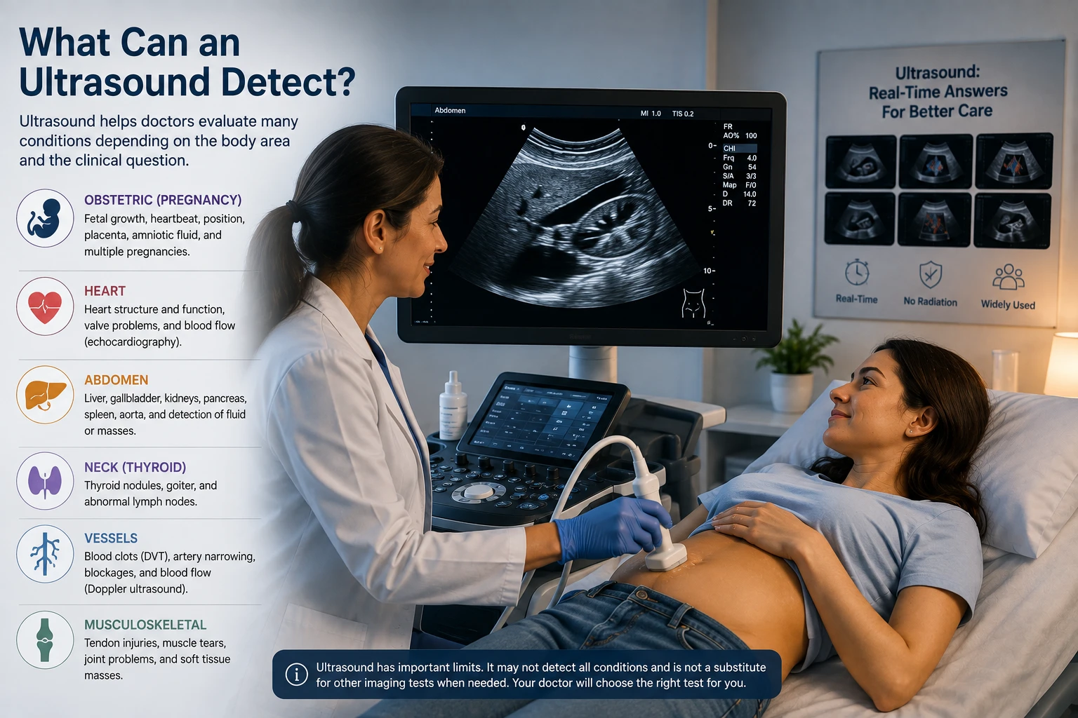
If your clinician ordered an ultrasound, you may wonder what the test can actually show. Diagnostic ultrasound excels at real-time pictures of many soft tissues, fluid collections, and blood flow when the sound beam has a clear path — but it cannot answer every medical question. This article walks through typical uses by body area, explains common blind spots such as bowel gas and bone, and describes when CT or MRI may be recommended instead. For what the test is and how it works at a high level, start with what is an ultrasound.
What Can an Ultrasound Detect?
There is no single list that applies to every patient. Detection depends on why the scan was ordered, which protocol was used, and how well sound travels through your body on that day. In general, diagnostic ultrasound helps clinicians evaluate structures that reflect sound waves well — organs, many soft-tissue masses, fluid, and moving blood when Doppler is used.
Professional practice parameters published by the American Institute of Ultrasound in Medicine define appropriate clinical indications for specialty exams (abdomen, pelvis, obstetrics, vascular, thyroid, and others). Those documents are written for clinicians; for patients, the takeaway is that your order matches a recognized indication rather than a generic “full body scan.”
A normal ultrasound does not automatically mean nothing is wrong if symptoms or other tests suggest otherwise. Follow-up imaging or clinical re-evaluation may still be needed.
Abdominal and Pelvic Ultrasound
Abdominal ultrasound commonly helps assess the liver, gallbladder, bile ducts, pancreas region, kidneys, spleen, and free fluid in the abdomen. Gallstones, cysts, obstructive patterns, and some masses may be visible when bowel gas does not block the view.
Gas in the stomach and intestines reflects sound strongly and can hide deeper structures — a long-standing limitation of sonography. That is one reason a clear answer about every abdominal organ is not guaranteed from one study.
Pelvic ultrasound — transabdominal or transvaginal — can evaluate the uterus, ovaries, and related structures for many gynecologic questions. Endometrial thickness, ovarian cysts, and some masses are frequent indications. Preparation such as fasting or bladder filling is exam-specific; see can you eat before an ultrasound rather than guessing.
Heart and Blood Vessels
Echocardiography (cardiac ultrasound) shows heart chamber size, valve motion, and pump function in real time — a different workflow from abdominal scanning but the same sound-based technology. Vascular ultrasound with Doppler can evaluate blood flow in veins and arteries for selected questions such as deep vein thrombosis screening in appropriate settings or stenosis follow-up.
Point-of-care ultrasound protocols in emergency and critical care include focused cardiac and vascular applications, but performance depends on operator training and the narrow question being asked. A brief bedside study is not the same as a comprehensive echocardiogram performed in a dedicated lab.
Pregnancy and Fetal Imaging
Obstetric ultrasound under multisociety practice parameters supports medically indicated tasks: confirming intrauterine pregnancy, estimating gestational age, assessing fetal growth, evaluating anatomy when clinically appropriate, and examining placenta and amniotic fluid. It is central to modern prenatal care when ordered for those purposes.
Ultrasound is not a complete genetic test. It cannot detect all chromosomal conditions or every structural anomaly. Abnormal screening blood tests or family history may lead to amniocentesis, cell-free DNA testing, or other pathways beyond imaging alone.
Elective 3D facial imaging for souvenirs is a separate topic from medically indicated detection; see what is 3D ultrasound for modality context.
Thyroid, Breast, and Soft Tissue
Ultrasound is frequently used to characterize thyroid nodules — size, composition, and suspicious features — often guiding whether fine-needle aspiration is needed. Breast ultrasound supplements mammography for some findings (for example evaluating a palpable lump or clarifying mammographic abnormalities) but does not replace screening mammography where that is recommended.
Superficial soft tissues, including some musculoskeletal structures, can be imaged when the target is close to the skin. Deep or bone-centered questions may require MRI or CT instead.
What Ultrasound Cannot Reliably Show
Ultrasound is limited by bone (sound does not penetrate well), air (bowel, lung interfaces), depth, body habitus, and patient movement. It is not the primary tool for detailed brain imaging in adults or for staging many cancers across the body.
Performance differs between a focused point-of-care scan and a comprehensive departmental exam. If a critical question remains unanswered, your team may recommend another modality. The American College of Radiology Appropriateness Criteria framework helps clinicians match imaging to clinical scenarios — without implying one test fits all.
Cancer detection questions are common online. Ultrasound may reveal suspicious masses in some organs but cannot guarantee that all cancers are absent. A planned Learning Center article on whether ultrasound can miss cancer will go deeper; until then, follow your clinician’s follow-up plan.
When Your Doctor May Order CT or MRI Instead
CT and MRI often provide clearer cross-sectional detail of deep structures, the brain, bones, and some soft-tissue contrasts that ultrasound cannot resolve when the acoustic window is poor. CT uses ionizing radiation; MRI does not — see does ultrasound use radiation and understanding MRI scans for modality differences.
Choosing ultrasound first is sometimes about real-time guidance, portability, or avoiding ionizing exposure when the question can be answered sonographically. Choosing CT or MRI is sometimes about resolution, staging, or anatomy hidden by gas and bone. The decision is clinical, not a ranking of “better” tests in general.
Key Takeaways
- There is no single list that applies to every patient.
- Abdominal ultrasound commonly helps assess the liver, gallbladder, bile ducts, pancreas region, kidneys, spleen, and free fluid in the abdomen.
- Echocardiography (cardiac ultrasound) shows heart chamber size, valve motion, and pump function in real time — a different workflow from abdominal scanning but the same sound-based technology.
- Obstetric ultrasound under multisociety practice parameters supports medically indicated tasks: confirming intrauterine pregnancy, estimating gestational age, assessing fetal growth, evaluating anatomy when clinically appropriate, and examining placenta and amniotic fluid.
- Ultrasound is frequently used to characterize thyroid nodules — size, composition, and suspicious features — often guiding whether fine-needle aspiration is needed.
Frequently Asked Questions
Can ultrasound detect cancer?
Ultrasound can sometimes show suspicious masses in organs it images well, but it cannot rule out all cancers and is not a universal cancer screening test. Follow-up depends on the finding and your history.
Can ultrasound see the brain?
In adults, brain detail is usually evaluated with CT or MRI, not standard transcranial ultrasound. Neonatal and focused applications exist in specialized care.
Can ultrasound find appendicitis?
Appendicitis may be suggested on ultrasound in some patients, especially children, but results vary. CT or other workup may be used when ultrasound is inconclusive.
Does a normal ultrasound mean nothing is wrong?
Not always. A normal study is reassuring for the question that was asked, but it does not exclude every condition — especially if symptoms persist or pretest suspicion is high.
Can ultrasound detect blood clots?
Venous ultrasound with Doppler is commonly used to evaluate suspected deep vein thrombosis in appropriate clinical settings when ordered.
Can ultrasound detect pneumonia?
Lung ultrasound can support diagnosis of some pulmonary conditions in selected settings, but chest X-ray or CT may still be used depending on the case.
Related articles
Glossary · Frequently asked questions · All Learning Center articles