How Many Bones Are in the Adult Human Body?
206 named bones — down from about 270 at birth as growth plates fuse during childhood and adolescence.
An adult human skeleton typically has 206 bones, fused from roughly 270 separate elements present early in development.
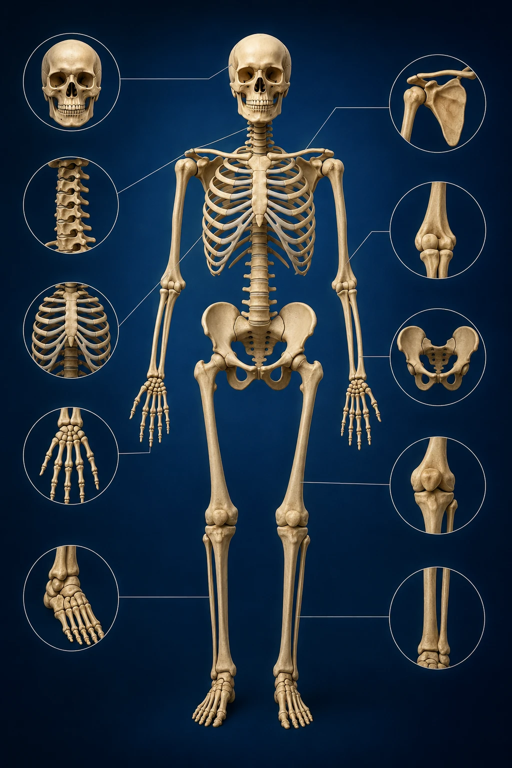
Babies are born with more separate bone pieces than adults have. As children grow, many bones fuse — especially in the skull and spine — leaving 206 bones in a typical adult skeleton.
From 270 to 206
Fontanelles ('soft spots') allow skull bones to overlap during birth; sutures fuse over the first years of life.
Growth plates in long bones close in the teens and twenties, completing length growth and reducing countable separate elements.
Key Facts
The axial skeleton (skull, spine, rib cage) protects brain, spinal cord, and heart-lungs; the appendicular skeleton includes arms, legs, shoulders, and pelvis.
Bone is living tissue that remodels with weight-bearing exercise and adequate calcium and vitamin D.
Fractures heal through a structured process that can be evaluated on X-ray — see our imaging primer linked below.
References
National Institute of Arthritis and Musculoskeletal and Skin Diseases — bone health (NIH).
Gray's Anatomy / standard anatomy references (peer-reviewed educational sources).
American Academy of Orthopaedic Surgeons — patient education on fractures.
Key Takeaways
- Fontanelles ('soft spots') allow skull bones to overlap during birth; sutures fuse over the first years of life.
- The axial skeleton (skull, spine, rib cage) protects brain, spinal cord, and heart-lungs; the appendicular skeleton includes arms, legs, shoulders, and pelvis.
Frequently Asked Questions
Do everyone have exactly 206 bones?
Small sesamoid bones (like extra kneecap variants) can add a few; 206 is the standard teaching count.
When does fusion finish?
Many fusions complete by the mid-twenties, though timing varies.
Why count bones on imaging?
Radiologists describe fractures and joint alignment using named bones; CT and X-ray map injuries to those structures.
Related articles
Glossary · Frequently asked questions · All Learning Center articles