3D vs 4D Ultrasound: What's the Difference?
3D ultrasound is a still volumetric snapshot; 4D adds the dimension of time. Here's why that distinction matters for a printed keepsake.
3D ultrasound captures one static volumetric snapshot of anatomy at a single moment, while 4D ultrasound plays a rapid sequence of those volumes to show live motion over time. For a physical keepsake, only one clean, well-positioned volume — pulled from either a 3D scan or a single 4D frame — is actually needed.
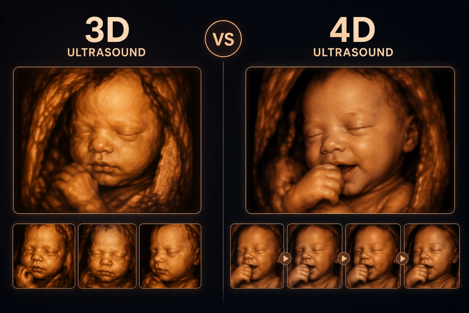
3D and 4D ultrasound are often used interchangeably in casual conversation, but they describe two related, distinct capabilities of the same underlying technology.
3D: A Still Volumetric Snapshot
3D ultrasound captures a single volumetric dataset at one moment in time — essentially a static 'sculpture' of the anatomy as it existed during that brief acquisition window.
This single volume is what contains the depth information needed to generate a 3D-printed surface.
4D: Adding the Dimension of Time
4D ultrasound is simply a rapid sequence of 3D volumes played back in succession, producing a live-motion effect — you can watch the fetus yawn, stretch, or move in near real time.
The fourth 'dimension' is time; the underlying spatial data at any single frame is still a 3D volume.
Which One Do You Need for a Keepsake?
For a physical print, we need one clean, well-positioned static volume rather than a video clip — essentially a single excellent frame pulled from either a 3D capture or a 4D sequence.
A 4D scan often gives more opportunities to catch a favorable moment, since dozens of volumes are captured per second rather than one.
Exporting the Right Data
If your clinic performed a 4D scan, ask whether individual volume frames or the full raw dataset can be exported, rather than only a compressed video file.
A compressed video export can lose the depth precision needed for a clean print, so raw or DICOM volume exports are strongly preferred.
Key Takeaways
- 3D ultrasound captures a single volumetric dataset at one moment in time — essentially a static 'sculpture' of the anatomy as it existed during that brief acquisition window.
- 4D ultrasound is simply a rapid sequence of 3D volumes played back in succession, producing a live-motion effect — you can watch the fetus yawn, stretch, or move in near real time.
- For a physical print, we need one clean, well-positioned static volume rather than a video clip — essentially a single excellent frame pulled from either a 3D capture or a 4D sequence.
- If your clinic performed a 4D scan, ask whether individual volume frames or the full raw dataset can be exported, rather than only a compressed video file.
Frequently Asked Questions
Do I need a 4D scan specifically for a keepsake?
No — a single high-quality 3D volume is sufficient. 4D simply gives more frames to choose from.
Can you print from a 4D video file?
We prefer raw volume or DICOM exports. Heavily compressed video files often lack the depth precision needed for a clean print.
Related articles
Glossary · Frequently asked questions · All Learning Center articles