What Is 3D Ultrasound?
A plain-language explanation of how volumetric ultrasound works and how it differs from a standard 2D scan.
3D ultrasound reconstructs many rapid 2D slices into a single volumetric dataset, letting anatomy be viewed from multiple angles instead of one flat plane. Clinically it helps evaluate structures like the fetal face or spine; the same volume data, when of sufficient quality, can also be converted into a physical 3D-printed keepsake.
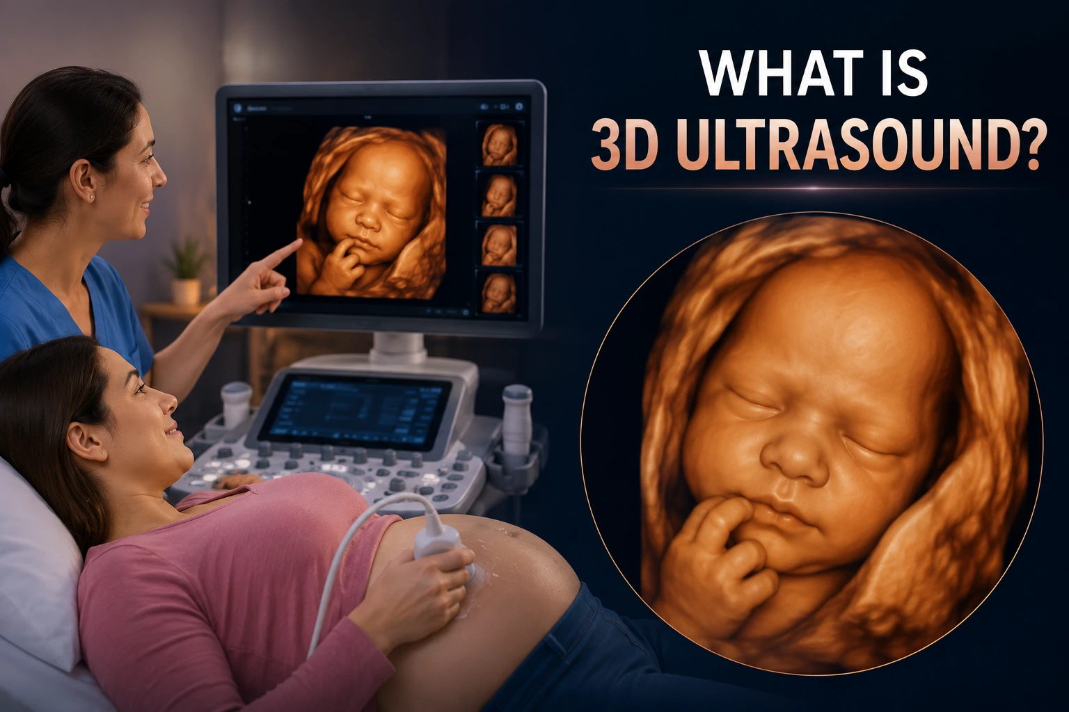
3D ultrasound is one of the most widely recognized imaging technologies in prenatal care, but many people aren't sure exactly how it differs from the 2D scans most of us picture when we think of an ultrasound.
From 2D Slices to a 3D Volume
A conventional ultrasound produces a single flat, grayscale cross-section — a 2D slice through the body at a given moment. 3D ultrasound captures many of these slices in rapid succession across a small volume of tissue.
Specialized software then reconstructs those slices into a single three-dimensional dataset, allowing the anatomy to be viewed from multiple angles rather than one flat plane.
How the Machine Builds the Image
Most modern 3D-capable ultrasound systems use a matrix-array or mechanically swept probe that automatically captures a series of closely spaced 2D slices over a short scanning window.
The system's onboard processor stitches these slices together using known geometric relationships between them, producing the surface-rendered image commonly associated with 3D ultrasound.
Clinical Uses vs. Keepsake Uses
Clinically, 3D ultrasound is used to evaluate structures like the fetal face, spine, and certain anomalies with more spatial context than 2D alone provides.
The same underlying volume data — when of sufficient quality — can separately be used to generate a physical 3D-printed keepsake, which is a non-diagnostic, commemorative use of the imaging.
What You Receive After Your Scan
Depending on the clinic and equipment, you may receive printed images, a video clip, or in some cases an exportable DICOM or vendor-specific 3D file.
Only the volumetric export — not a printed photo — contains the depth information needed to create a 3D-printed model.
Key Takeaways
- A conventional ultrasound produces a single flat, grayscale cross-section — a 2D slice through the body at a given moment.
- Most modern 3D-capable ultrasound systems use a matrix-array or mechanically swept probe that automatically captures a series of closely spaced 2D slices over a short scanning window.
- Clinically, 3D ultrasound is used to evaluate structures like the fetal face, spine, and certain anomalies with more spatial context than 2D alone provides.
- Depending on the clinic and equipment, you may receive printed images, a video clip, or in some cases an exportable DICOM or vendor-specific 3D file.
Frequently Asked Questions
Is 3D ultrasound the same as a regular ultrasound?
It uses the same underlying technology but captures and reconstructs many slices into a single three-dimensional volume, rather than one flat image.
Can any ultrasound clinic provide files suitable for 3D printing?
Not every clinic offers volumetric export. Ask specifically whether a 3D/4D volume or DICOM file can be exported, not just printed photos.
Related articles
Related images
Glossary · Frequently asked questions · All Learning Center articles