How Modern Ultrasound Machines Create Volume Data
A closer look at the hardware and processing pipeline that turns sound waves into a 3D-printable dataset.
Modern 3D-capable ultrasound probes use a matrix array or a mechanically swept element to capture many closely spaced 2D slices across a small volume in a fraction of a second. Onboard processors then stitch those slices into a continuous 3D dataset, which — when exported as a raw or DICOM volume — can be 3D printed.
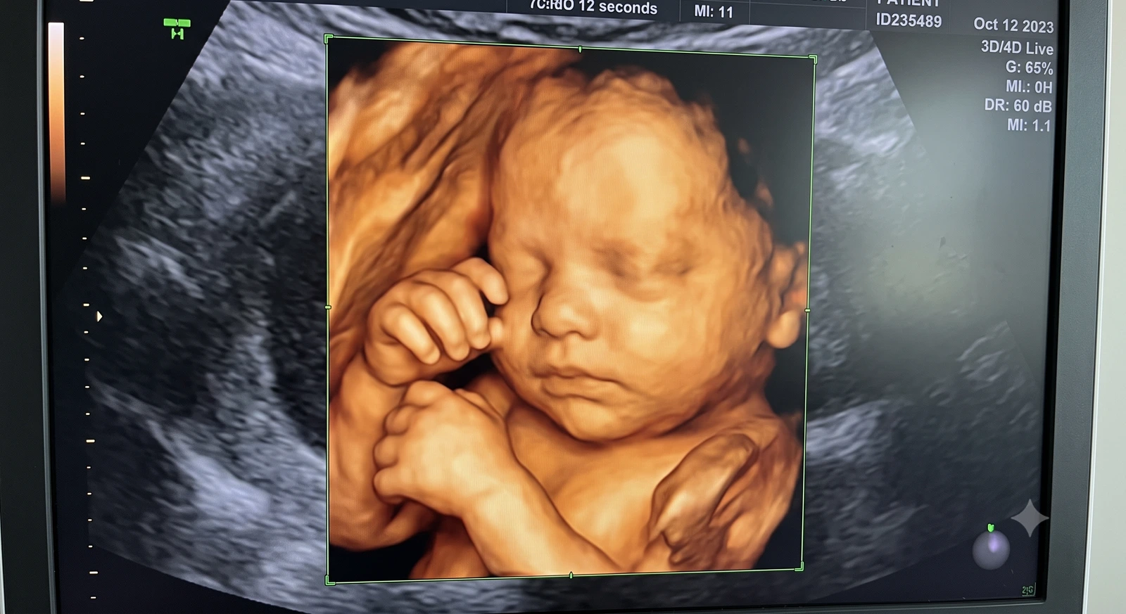
Behind every 3D ultrasound keepsake is a surprisingly sophisticated hardware and software pipeline. Here's a technical, but accessible, look at how it works.
From Piezoelectric Crystals to Pixels
Ultrasound transducers contain piezoelectric elements that convert electrical pulses into sound waves and, on return, convert reflected sound back into electrical signals.
The time delay and strength of each returning echo tells the system how far away, and how dense, the tissue that produced it was — the basic building block of every ultrasound image.
The Matrix Array and Mechanical Sweep
3D-capable probes use either a two-dimensional matrix array of elements, which can electronically steer the sound beam across a volume without moving parts, or a mechanically swept single-array probe that physically tilts through a small arc.
Both approaches achieve the same goal: acquiring many closely spaced 2D slices across a small three-dimensional region in a fraction of a second.
Reconstructing the Volume
Onboard processors combine the individual slices using known spatial relationships between them, interpolating between slices to build a continuous 3D dataset rather than a stack of disconnected images.
This reconstructed volume can then be rendered as a surface image on the machine's screen in real time, or exported for further processing.
Exporting Volumes for Downstream Use
For 3D printing, the raw or DICOM-formatted volume — not a rendered screenshot — is what preserves the depth information needed for mesh generation.
This is why we ask families to request a volume or DICOM export from their clinic rather than relying on a printed photo or compressed video alone.
Key Takeaways
- Ultrasound transducers contain piezoelectric elements that convert electrical pulses into sound waves and, on return, convert reflected sound back into electrical signals.
- 3D-capable probes use either a two-dimensional matrix array of elements, which can electronically steer the sound beam across a volume without moving parts, or a mechanically swept single-array probe that physically tilts through a small arc.
- Onboard processors combine the individual slices using known spatial relationships between them, interpolating between slices to build a continuous 3D dataset rather than a stack of disconnected images.
- For 3D printing, the raw or DICOM-formatted volume — not a rendered screenshot — is what preserves the depth information needed for mesh generation.
Frequently Asked Questions
What file format should I request from my ultrasound clinic?
A DICOM volume export is ideal. Some machines also produce vendor-specific 3D file formats that can work as well.
Can a screenshot of a 3D ultrasound be used for printing?
No. A screenshot is a flattened 2D image and does not retain the underlying depth data needed to build an accurate physical surface.
Related articles
Related images
Glossary · Frequently asked questions · All Learning Center articles