What Is Medical Image Segmentation?
The technical process of isolating anatomy within a scan — and why segmentation quality determines model quality.
Segmentation is the process of classifying each voxel in a medical scan as belonging to a specific structure — bone, an organ, or a fetal surface — versus everything around it, producing an isolated digital shape ready for mesh conversion. Every downstream step depends on segmentation accuracy, which is why trained human review remains standard.
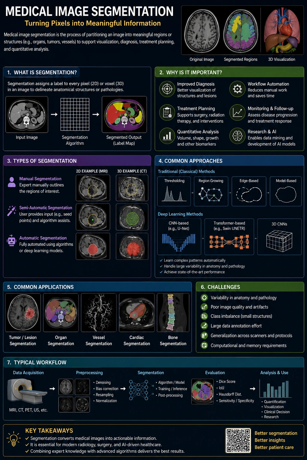
Segmentation is arguably the single most important — and most misunderstood — step in the medical 3D printing pipeline. It's where imaging data becomes a specific, printable anatomical structure.
Defining Segmentation
Segmentation is the process of classifying each voxel (3D pixel) in a scan as belonging to a specific structure — bone, a particular organ, a tumor, or fetal surface — versus everything else.
The output is a clean, isolated digital representation of just the anatomy of interest, ready to be converted into a printable mesh.
Manual, Semi-Automatic, and AI-Assisted Methods
Manual segmentation involves a trained technician tracing structure boundaries slice by slice — accurate but time-intensive. Semi-automatic tools use density thresholding to speed this up, with human correction of edge cases.
AI-assisted segmentation uses trained models to propose boundaries automatically, which can significantly accelerate the process while still requiring human review for accuracy and safety.
Why Segmentation Quality Determines Model Quality
Every downstream step — mesh generation, printing, finishing — depends entirely on the accuracy of the initial segmentation. An error introduced here propagates through the entire pipeline.
This is why segmentation is treated as a quality-critical step requiring trained review rather than a fully automated, unchecked process.
Segmentation at LifePrint 3D
Our team combines specialized segmentation software with manual review of every case, checking the isolated structure against the original imaging before it proceeds to mesh preparation.
For ultrasound keepsakes specifically, this means confirming that fluid, cord, and limb shadowing have been appropriately distinguished from true fetal surface.
Key Takeaways
- Segmentation is the process of classifying each voxel (3D pixel) in a scan as belonging to a specific structure — bone, a particular organ, a tumor, or fetal surface — versus everything else.
- Manual segmentation involves a trained technician tracing structure boundaries slice by slice — accurate but time-intensive.
- Every downstream step — mesh generation, printing, finishing — depends entirely on the accuracy of the initial segmentation.
- Our team combines specialized segmentation software with manual review of every case, checking the isolated structure against the original imaging before it proceeds to mesh preparation.
Frequently Asked Questions
Is segmentation fully automated?
Automated and AI-assisted tools accelerate the process, but trained human review remains a standard part of a quality segmentation workflow.
Can segmentation errors be caught before printing?
Yes — reviewing the segmented structure against the source imaging before mesh generation is the primary way errors are identified and corrected.
Related articles
Glossary · Frequently asked questions · All Learning Center articles