2D vs 3D vs 4D Ultrasound Pictures: What You Usually Leave With
The numbers on the appointment are not the same as the souvenirs in the bag. Here is what 2D printouts, 3D stills, 4D video, and volume files usually mean as take-home media.
2D visits typically send you home with grayscale stills or thermal printouts of single slices. 3D visits often add surface-looking stills — a statue-like view on a flat file. 4D visits usually add a short cine or video of motion. Those files are memories of the appointment, not automatically a saved 3D dataset. A volume file, when one exists and can be copied, is spatial data rather than a snapshot — pictures of a shape in space, and sometimes that shape can later become something you can hold.
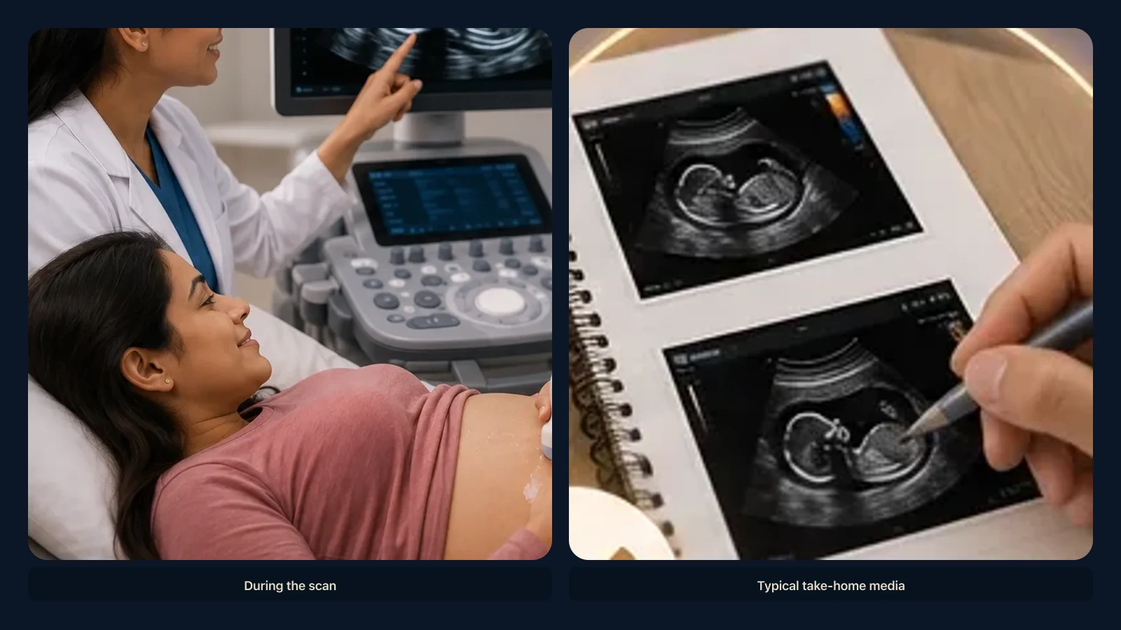
After a prenatal ultrasound, most families do not leave with “2D,” “3D,” or “4D” as objects in a bag. They leave with media: a strip of thermal paper, a few stills on a phone, maybe a short video they will watch on the ride home. The numbers on the appointment describe how the machine built the pictures. The souvenirs describe what was easy to print, email, or compress.
The Envelope Is Not the Exam
Clinics package pictures in many ways: glossy prints, a patient portal PDF, JPEGs on a USB stick, a vendor app, or a message from a boutique studio. The wrapper does not tell you the acquisition mode.
Staff may say they did 4D while the envelope holds six stills and one movie. Another office may call it a standard ultrasound and hand you a thick stack of 2D slices that feel like the whole story of the hour.
A useful habit is to separate what you watched in the room, what you took home today, and what, if anything, was stored on the clinic system that is not in the bag. This article is about the second question. It is not a guide to which exam anyone should have, what a finding means, or how to date a pregnancy.
2D: The Classic Printout
A 2D image is a slice — a thin plane through the body at one moment. On paper it looks grainy, black, white, and gray. Bright edges appear where sound bounced strongly; darker pools appear where it did not.
That is the fridge photo most people mean by ultrasound picture: a profile, a round midsection, a cluster of bones someone in the room pointed to. What you usually leave with is stills.
Some clinics also save 2D cine loops, so you might get a short clip even without a 3D or 4D session. That clip is still a movie of slices, not a spinning miniature. 2D printouts are excellent at being recognizable later. The limitation is honest: a slice is not a map of a whole surface.
3D: A Still of a Shape
3D ultrasound packs many slices into a small block of space, then paints a surface so a face or a limb can look more like a miniature sculpture on a screen. The doll-like or softly lit look is a rendering — shading chosen so depth reads on a monitor — not a color photograph of skin in room light.
What you usually leave with from a 3D session is still a picture: a JPEG, a print, sometimes a sheet of best views. The paper does not contain the volume. It is a postcard of one angle, the way a photo of a statue is not the statue.
It is easy to assume the pretty still is the 3D. It is a picture of a volume. If the visit saved the volume itself, that file typically lives on the scanner or in a clinical archive, not automatically in the souvenir packet. For how 3D and 4D differ as machine modes, see our article on 3D vs 4D ultrasound. That page is the modality explainer; this one stays on what tends to go in the bag.
4D: The Short Video
4D is 3D over time: volumes updating fast enough that a yawn or a stretch reads as live motion. The souvenir is often a video — an MP4, a clip in a clinic app, sometimes an older DVD. Families remember the motion more than any single frame.
Compression that makes a phone-friendly movie is not the same as keeping every volume in the sequence. A 4D video is usually a recording of the experience, or a compressed export, not a stack of full 3D frames you can later reopen as spatial data. If you only have the movie, you have the memory of movement. You do not automatically have the underlying volumes.
What Usually Does Not Go Home Unless Someone Asks
Many hospitals store the diagnostic study in a medical archive, often DICOM on a PACS. Families may see selected images in a portal and never receive the full study unless they request a copy through the usual health-records path.
Boutique 3D and 4D studios may give lots of pretty stills and a video and still not export a raw volume. A volume — or a vendor volume file — is a different take-home item from a printout. It is closer to a digital sculpture sitting in a file than to a snapshot. Not every visit captures one, and not every captured volume is saved in a form a family can copy.
Practical steps for requesting copies of imaging in general are covered in our article on how to get a copy of your scan images. This page does not walk through clinic workflows or file formats in depth.
Pictures of a Volume — and Sometimes Something You Can Hold
Even a familiar 2D slice is a picture of anatomy in space: a cut through a three-dimensional body. A 3D or 4D acquisition makes that spatial fact obvious. The screen shows a shape with depth, even when you only take home a flat photo of that shape.
Sometimes, when a suitable volume was stored, that same surface can later be turned into a small physical likeness you can turn in your hands. That is optional and downstream of the visit. It is not a reason to treat stills and video as incomplete. The printouts and the cine clip are the usual souvenirs; they do not need to be upgraded for the appointment to have done its job.
A handheld piece, if one is ever made, will not match the gray palette of a 2D printout or the cinematic lighting of a 4D video. Those are different ways of looking at a captured shape — a picture, a moving picture, and, more rarely, an object.
A Plain List of Typical Take-Homes
2D stills and printouts are slice pictures, the most common fridge media. 3D stills are one framed view of a reconstructed surface, still a flat file. 4D or cine video is the motion souvenir, usually compressed for phones and apps. Volume or archive data is the spatial dataset, if it was saved and if you can obtain a copy — not the default party favor.
Packets mix. A session described as 3D or 4D often means stills plus video. A clinical 2D exam might include more slices than a boutique keepsake visit, and fewer statue-like views.
How to Talk About It at the Desk
These are logistics questions, not medical ones. Ask whether you will get paper prints, portal images, or both; whether there is a video clip and in what format; and, if the visit was described as 3D or 4D, whether the take-home files are stills and video only or whether a volume was saved.
Staff may not be able to export a volume the same day. That is a software and policy question, not a verdict on how nice the pictures look.
What This Article Does Not Cover
This is visual literacy — how families talk about the pictures they leave with. It is not advice about which ultrasound you need, whether a picture looks normal, how to interpret a report, or anything about determining sex.
We are not listing prices or products. If you have questions about your own exam, ask the clinicians who performed or ordered it. Nearby topics include what 3D ultrasound is, how modern ultrasound creates volume data, and what an ultrasound is in general.
Key Takeaways
- Clinics package pictures in many ways: glossy prints, a patient portal PDF, JPEGs on a USB stick, a vendor app, or a message from a boutique studio.
- A 2D image is a slice — a thin plane through the body at one moment.
- 3D ultrasound packs many slices into a small block of space, then paints a surface so a face or a limb can look more like a miniature sculpture on a screen.
- 4D is 3D over time: volumes updating fast enough that a yawn or a stretch reads as live motion.
- Many hospitals store the diagnostic study in a medical archive, often DICOM on a PACS.
Frequently Asked Questions
Is a 3D ultrasound photo the same as 3D data?
No. The photo is one rendered view. The volume is the block of space the machine used to draw that view.
If I have a 4D video, do I have 4D volumes?
Usually you have a movie. The individual volumes may still sit only on the scanner or in the clinic archive.
Why does my 3D picture look a little like a doll?
Surface rendering uses shading to suggest depth on a flat screen. It is not a daylight photograph of the baby.
Can I tell 2D from 3D from the printout alone?
Often, yes: 2D looks like a grainy slice; 3D stills look more like a small statue. Mixed packets are common. A report, disc label, or the person who scanned you is more reliable than guessing from a cropped social post.
Do I need a volume file for memories?
No. Stills and clips are the memories most families keep. A volume is a different kind of file for a different kind of later use — including, when the data allow, a shape you can hold.
Related articles
Glossary · Frequently asked questions · All Learning Center articles