Patient-Specific Anatomical Models Explained
What makes a model 'patient-specific,' how accuracy is preserved, and where these models are used in care and education.
A patient-specific model is built directly from one person's own CT, MRI, or ultrasound data, preserving their actual anatomical variation rather than a generic textbook average. Accuracy depends on careful segmentation, minimal mesh smoothing, and quality review at every pipeline stage, since deviations at any step compound in the finished model.
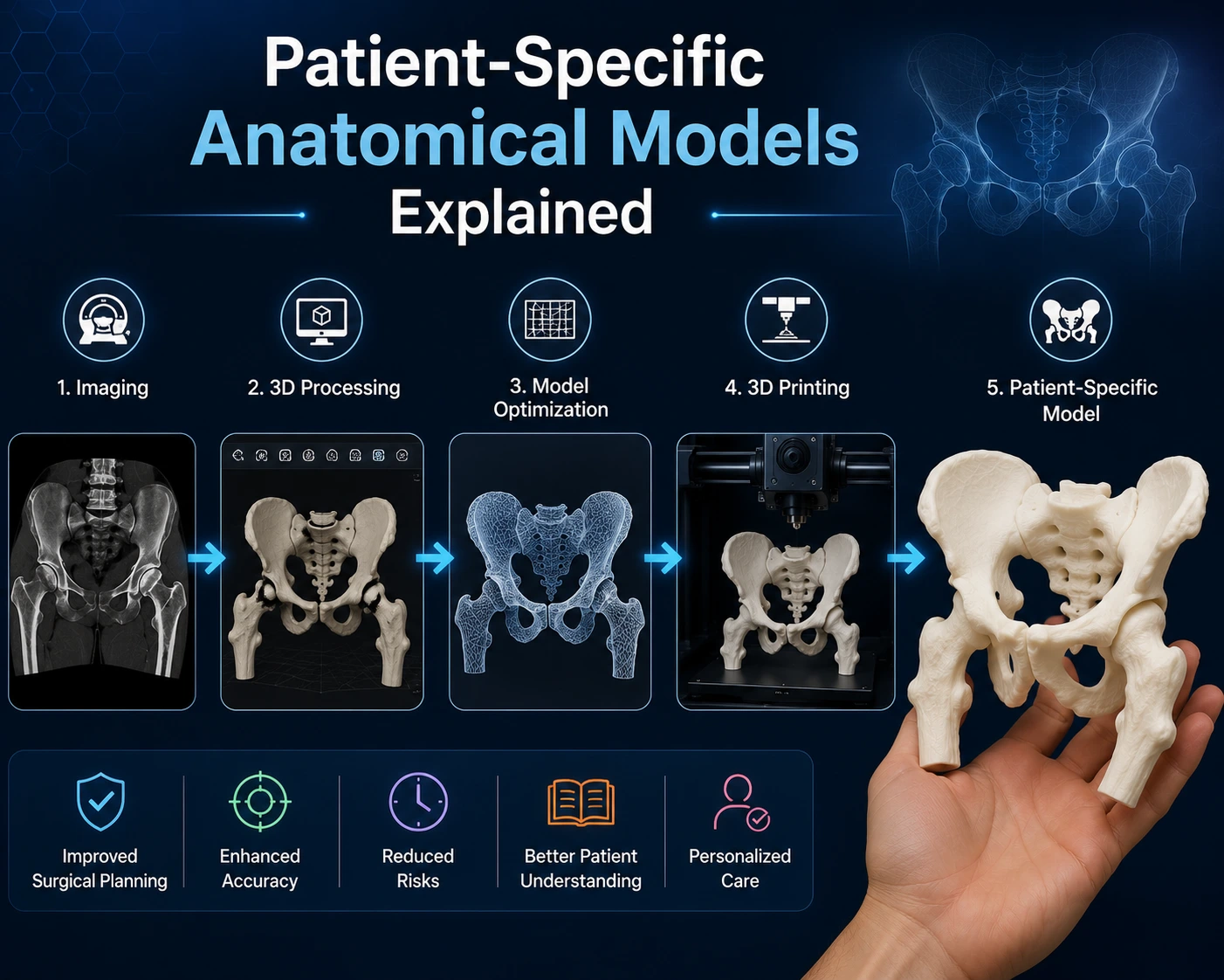
The term 'patient-specific' distinguishes a custom anatomical model built from an individual's own imaging from a generic, off-the-shelf teaching model. That distinction matters more than it might seem.
What Makes a Model 'Patient-Specific'
A patient-specific model is built directly from one individual's CT, MRI, or ultrasound data, preserving their unique anatomical variations rather than representing a textbook 'average' anatomy.
This means the model can reflect an individual's actual vessel branching pattern, tumor shape, or fracture geometry, rather than a generalized approximation.
How Accuracy Is Preserved Through the Pipeline
Accuracy depends on maintaining fidelity at every step: careful segmentation that follows the true tissue boundaries, minimal mesh smoothing that could erase real detail, and printing at a scale and resolution appropriate to the anatomy.
Deviations at any stage compound, which is why quality review against the source imaging is a standard part of a rigorous workflow.
Common Uses in Care and Education
Patient-specific models support pre-surgical rehearsal, resident and student training on real (de-identified or consented) anatomy, and clearer conversations between clinicians and patients about a specific condition.
Fetal keepsakes are a distinct but related application — a patient-specific model created for commemorative rather than clinical purposes.
Limitations to Understand
A patient-specific model reflects the anatomy captured at the moment of imaging; it is not updated in real time and does not replace ongoing diagnostic imaging.
It is also, importantly, a representation derived from imaging data — not the imaging itself — and carries whatever limitations were present in the original scan.
Key Takeaways
- A patient-specific model is built directly from one individual's CT, MRI, or ultrasound data, preserving their unique anatomical variations rather than representing a textbook 'average' anatomy.
- Accuracy depends on maintaining fidelity at every step: careful segmentation that follows the true tissue boundaries, minimal mesh smoothing that could erase real detail, and printing at a scale and resolution appropriate to the anatomy.
- Patient-specific models support pre-surgical rehearsal, resident and student training on real (de-identified or consented) anatomy, and clearer conversations between clinicians and patients about a specific condition.
- A patient-specific model reflects the anatomy captured at the moment of imaging; it is not updated in real time and does not replace ongoing diagnostic imaging.
Frequently Asked Questions
Are patient-specific models used for diagnosis?
No. They are typically used for planning, education, and communication support alongside — not in place of — diagnostic imaging and clinical judgment.
How accurate is a patient-specific 3D printed model?
Accuracy depends on source imaging resolution and segmentation care, but well-executed models can closely reflect the true anatomical geometry captured in the scan.
Related articles
Glossary · Frequently asked questions · All Learning Center articles