Understanding CT Scans: How They Work
How computed tomography builds cross-sectional images from X-rays, and why it's well suited to 3D printing.
Computed tomography (CT) rotates an X-ray source and detector around the body, capturing many angled projections that are mathematically reconstructed into cross-sectional slices forming a 3D volume. Because bone and soft tissue differ sharply in density on CT, it is the most common imaging source for 3D printed skeletal and vascular models.
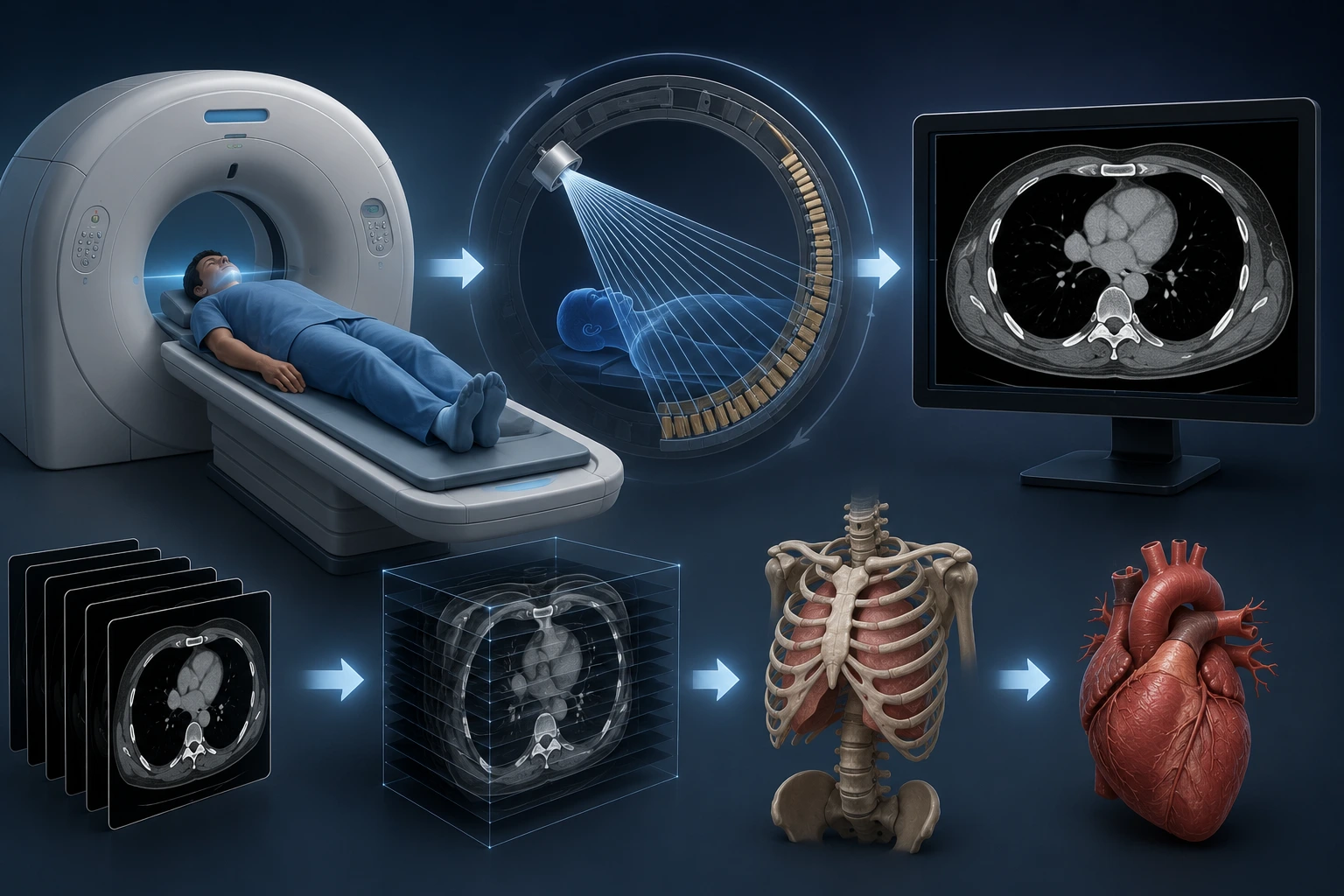
Computed tomography (CT) is one of the two most common imaging sources for medical 3D printing, alongside MRI. Understanding how it works clarifies why it produces such reliable 3D printing data.
X-Rays From Every Angle
A CT scanner rotates an X-ray source and detector array around the patient, capturing dozens to hundreds of individual X-ray projections from different angles in a single rotation.
Each projection measures how much X-ray energy is absorbed along that path, which varies by tissue density — bone absorbs far more than soft tissue or air.
From Raw Data to Cross-Sectional Slices
A mathematical reconstruction process (historically called 'back-projection' or more modern iterative methods) combines the many angled projections into a single cross-sectional slice image.
The scanner repeats this process while moving along the body, producing a full stack of slices that together form a three-dimensional volume, stored as a DICOM series.
Contrast and Resolution
CT is particularly good at distinguishing bone, calcified tissue, and — with the use of contrast agents — blood vessels, because these structures differ sharply in density from surrounding soft tissue.
Modern CT scanners can achieve sub-millimeter slice thickness, providing fine enough detail for precise anatomical modeling.
Why CT Is Ideal for 3D Printing
The strong density contrast between bone and soft tissue makes CT data relatively straightforward to segment for skeletal and vascular models compared to some other modalities.
This is why CT remains the most common source for surgical planning models, particularly for bone, vascular, and complex trauma anatomy.
Key Takeaways
- A CT scanner rotates an X-ray source and detector array around the patient, capturing dozens to hundreds of individual X-ray projections from different angles in a single rotation.
- A mathematical reconstruction process (historically called 'back-projection' or more modern iterative methods) combines the many angled projections into a single cross-sectional slice image.
- CT is particularly good at distinguishing bone, calcified tissue, and — with the use of contrast agents — blood vessels, because these structures differ sharply in density from surrounding soft tissue.
- The strong density contrast between bone and soft tissue makes CT data relatively straightforward to segment for skeletal and vascular models compared to some other modalities.
Frequently Asked Questions
Does CT use radiation?
Yes, CT uses ionizing X-ray radiation, unlike ultrasound or MRI. Dosing is determined by the ordering physician based on clinical necessity.
Is CT better than MRI for 3D printing?
It depends on the anatomy. CT typically excels for bone and vascular structures; MRI is often preferred for soft tissue and neural anatomy.
Related articles
Glossary · Frequently asked questions · All Learning Center articles