Understanding MRI Scans: How They Work
How magnetic resonance imaging creates detailed soft-tissue contrast without ionizing radiation.
MRI uses a strong magnetic field and radiofrequency pulses to align and then read signals from hydrogen atoms in the body, producing images without any ionizing radiation. Because different tissues realign at different rates, MRI offers finer soft-tissue contrast than CT, making it the preferred imaging source for brain and soft-tissue anatomical models.
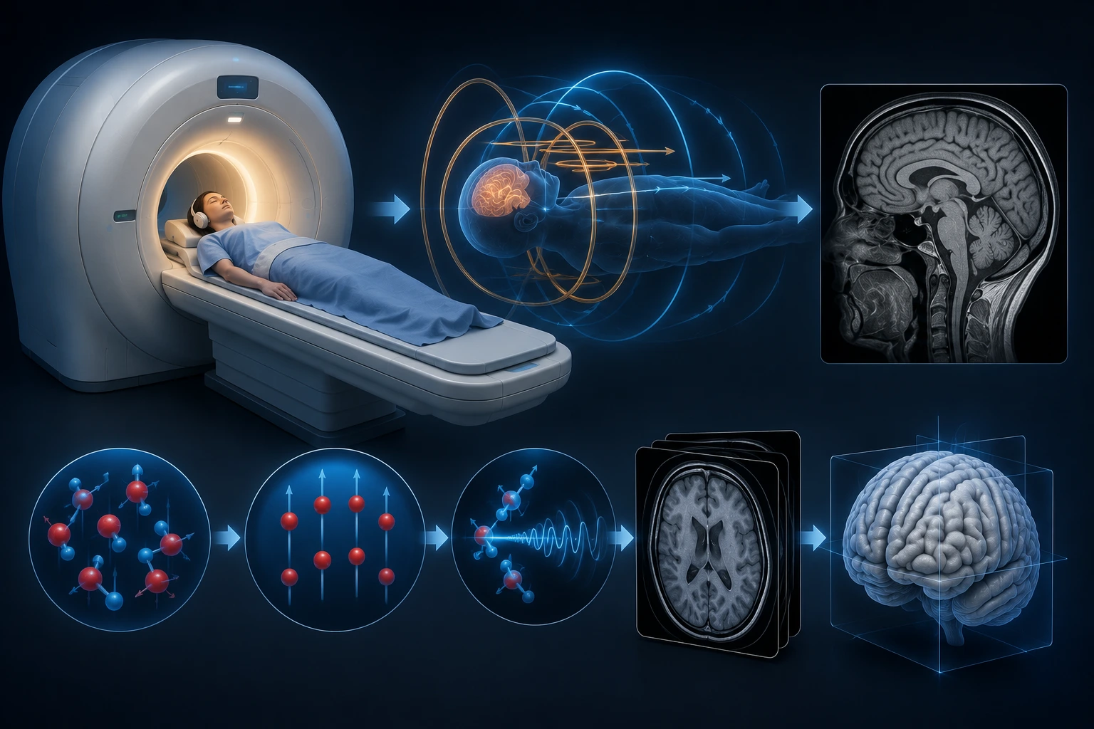
Magnetic resonance imaging (MRI) is prized in medicine for its exceptional soft-tissue contrast. Here's a plain-language look at the physics behind it and why it's valuable for anatomical modeling.
Magnetic Fields, Not Radiation
MRI uses a powerful magnetic field to briefly align hydrogen atoms (abundant in water and fat throughout the body), then applies radiofrequency pulses that knock them out of alignment.
As the atoms realign, they emit a faint radio signal that the scanner detects and uses to reconstruct an image — no ionizing radiation is involved at any point.
How Soft Tissue Contrast Is Created
Different tissues realign at different rates depending on their water and fat content, which is what allows MRI to distinguish gray matter from white matter, or a tumor from surrounding healthy tissue, with much finer soft-tissue contrast than CT.
By adjusting scan parameters (called sequences), radiologists can emphasize different tissue properties for different diagnostic questions.
Sequences and Slice Thickness
Common sequences include T1-weighted and T2-weighted imaging, each highlighting different tissue characteristics. Slice thickness and spacing vary by protocol and clinical indication.
For 3D printing purposes, thinner slices with less gap between them generally produce a smoother, more accurate reconstructed surface.
MRI's Role in Anatomical Modeling
MRI is often the preferred source for modeling brain structures, soft-tissue tumors, and other anatomy where CT's bone-and-vessel strength is less relevant.
Because MRI lacks ionizing radiation, it is also frequently used in pediatric and repeated-imaging contexts where radiation exposure is a greater concern.
Key Takeaways
- MRI uses a powerful magnetic field to briefly align hydrogen atoms (abundant in water and fat throughout the body), then applies radiofrequency pulses that knock them out of alignment.
- Different tissues realign at different rates depending on their water and fat content, which is what allows MRI to distinguish gray matter from white matter, or a tumor from surrounding healthy tissue, with much finer soft-tissue contrast than CT.
- Common sequences include T1-weighted and T2-weighted imaging, each highlighting different tissue characteristics.
- MRI is often the preferred source for modeling brain structures, soft-tissue tumors, and other anatomy where CT's bone-and-vessel strength is less relevant.
Frequently Asked Questions
Is MRI safer than CT?
MRI does not use ionizing radiation, which is an advantage in certain contexts, though it has its own considerations such as scan time and compatibility with implanted metal devices.
Can MRI data be used for fetal keepsakes?
Fetal keepsakes are created from ultrasound, not MRI. Fetal MRI is a separate, specialized clinical tool used only for specific medical indications.
Related articles
Glossary · Frequently asked questions · All Learning Center articles