When Is an X-Ray Enough for a Suspected Fracture?
Plain radiographs are often the first imaging test for suspected fractures. Patient-facing ACR summaries explain when X-ray is usually enough — and when CT or MRI may be considered next.
For many suspected fractures after injury, a bone X-ray is the usual first imaging test and is often sufficient when it clearly answers the clinical question. If fracture remains suspected despite a negative or unclear radiograph — a pattern discussed especially for hip injury after falls — MRI or CT may be considered next, based on clinical judgment and local protocols. Only your care team can decide the right sequence for you.
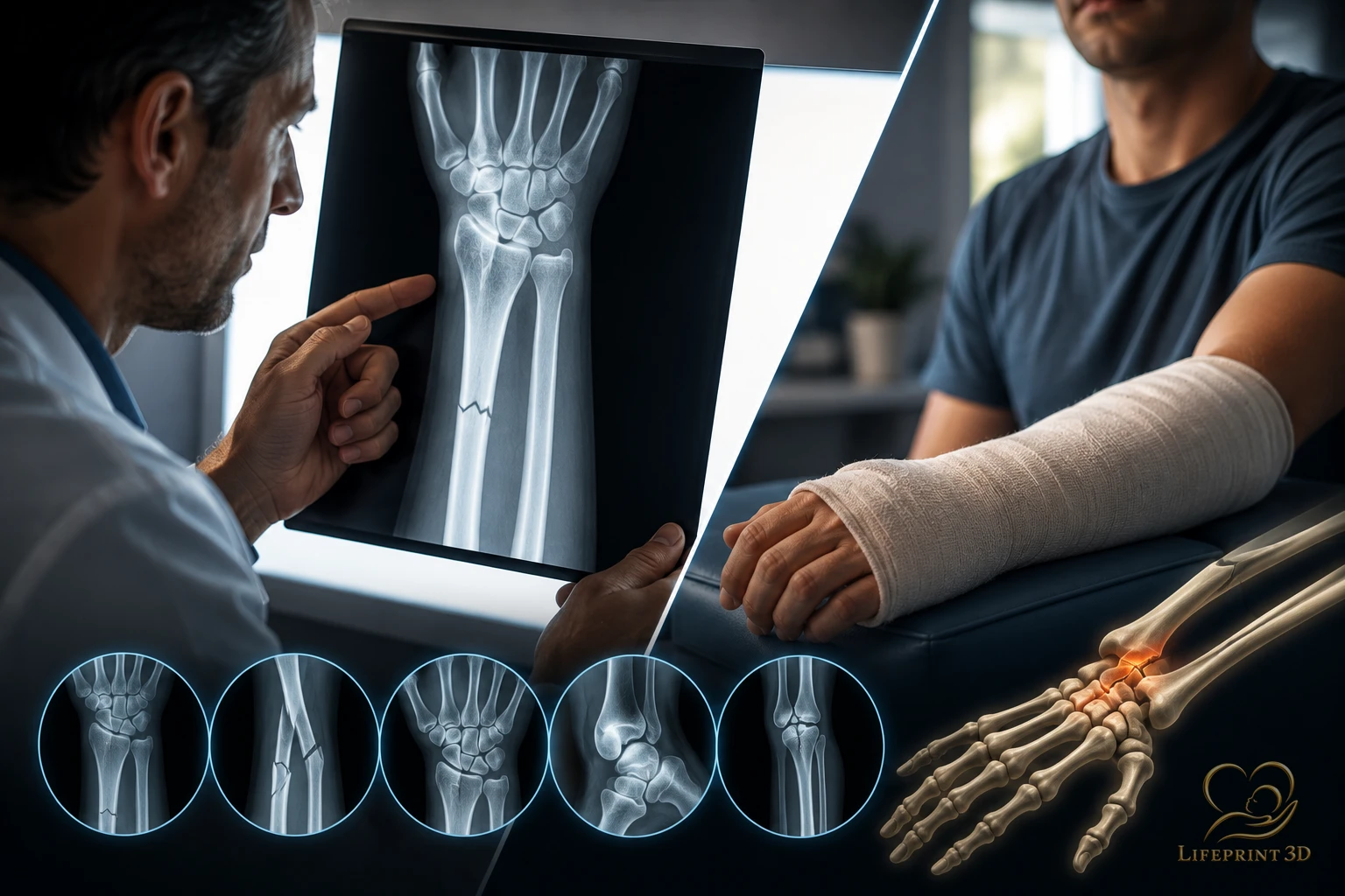
When a bone injury is suspected, clinicians frequently start with a bone X-ray because it is fast, widely available, and excellent for many fractures. This article explains that imaging pathway in educational terms: when radiographs are typically first-line, what they show well, and when additional CT or MRI may be discussed. It does not diagnose your injury or recommend casting, surgery, or other treatment.
Why Radiographs Are Often First
RadiologyInfo.org (ACR/RSNA patient education) explains that bone X-ray uses a small dose of ionizing radiation to produce pictures of bone and is commonly used to diagnose fractured bones or joint dislocation. It describes bone X-rays as among the fastest and easiest ways for a physician to view and assess bone fractures and joint abnormalities.
Because radiographs are quick and widely available in emergency and outpatient settings, they are a practical first look when the clinical question is primarily about bone alignment and cortical disruption.
When an X-Ray May Be Enough
An X-ray may be enough when the images adequately show the bones of interest, the fracture pattern is clear, and the study answers the clinician’s question about presence, alignment, and related bony injury. Follow-up radiographs are also commonly used later to assess healing alignment — again, as ordered clinically.
“Enough” is a clinical judgment, not a patient self-score. Soft-tissue injuries (ligaments, tendons, menisci) are often poorly shown on plain films, so a normal bone X-ray does not mean soft tissues are uninjured.
When CT or MRI May Enter the Conversation
RadiologyInfo notes limitations of bone X-ray: little information about muscles, tendons, or many joint soft tissues; MRI may be more useful for many soft-tissue injuries and can detect some subtle or occult fractures not visible on X-ray. CT is widely used in trauma evaluation and can depict complicated fractures, subtle fractures, or dislocations; in older adults with osteoporosis, a hip fracture may be clearer on CT than on a plain hip radiograph.
A RadiologyInfo summary of appropriateness for acute hip pain with suspected fracture states that radiography of the hip and pelvis is the most appropriate first imaging test after fall or minor trauma and can identify most hip fractures. When a fracture is not seen on X-ray but remains clinically suspected, MRI of the pelvis and hips without IV contrast is described as the next best imaging test; CT without IV contrast is also generally appropriate as a second test, especially when MRI is difficult.
Could Fracture CT Data Be Used for a 3D Model?
RadiologyInfo notes that for some complicated injuries, three-dimensional reconstructed CT images can be created from the CT dataset without additional radiation exposure, to help clinicians understand complex geometry. That is a digital reconstruction step inside radiology — distinct from ordering a consumer keepsake model.
Separately, peer-reviewed literature has studied physical patient-specific 3D-printed anatomical models for selected surgical planning and education uses. A systematic review of clinical 3D printing efficacy (PMID 29273650) maps a growing literature concentrated heavily in surgical contexts, while also showing that high-level outcome evidence is uneven. A 2024 systematic review on quality assurance of 3D-printed patient-specific anatomical models (PMID 38536566) emphasizes that segmentation, digital editing, and printing each contribute error — so a physical model is not an exact replica of anatomy.
LifePrint 3D works from imaging already obtained for clinical care and does not encourage anyone to get a CT or X-ray solely to make a model. If you already have a study, how to request a copy of the images is covered in how to get a copy of your own scan images, and the DICOM-to-model workflow is described in from DICOM to physical model.
References
RadiologyInfo.org (ACR/RSNA) — Bone X-ray (https://www.radiologyinfo.org/en/info/bonerad).
RadiologyInfo.org — Acute Hip Pain–Suspected Fracture appropriateness summary (https://www.radiologyinfo.org/en/info/acs-acute-hip-pain-suspected-fracture).
Jarraya M, et al. — Radiographically occult and subtle fractures: a pictorial review. Radiol Res Pract. 2013 (PMID 23577253; doi: 10.1155/2013/370169).
Diment LE, et al. — Clinical efficacy and effectiveness of 3D printing: a systematic review. BMJ Open. 2017 (PMID 29273650).
Systematic review — Quality assurance of 3D-printed patient specific anatomical models (PMID 38536566; doi: 10.1186/s41205-024-00210-5).
Key Takeaways
- RadiologyInfo.
- An X-ray may be enough when the images adequately show the bones of interest, the fracture pattern is clear, and the study answers the clinician’s question about presence, alignment, and related bony injury.
- RadiologyInfo notes limitations of bone X-ray: little information about muscles, tendons, or many joint soft tissues; MRI may be more useful for many soft-tissue injuries and can detect some subtle or occult fractures not visible on X-ray.
- RadiologyInfo notes that for some complicated injuries, three-dimensional reconstructed CT images can be created from the CT dataset without additional radiation exposure, to help clinicians understand complex geometry.
Frequently Asked Questions
If the X-ray is normal, is the bone definitely not broken?
Not always. Some fractures are radiographically occult. Clinicians may consider MRI or CT when suspicion remains high despite a negative X-ray — especially in certain sites such as the hip after a fall.
Does every fracture need a CT?
No. Many fractures are adequately characterized on radiographs. CT is discussed when detail about complex patterns, articular involvement, or unclear radiographs is needed.
Can soft-tissue injuries show on a bone X-ray?
Plain films primarily show bone. Ligament and tendon injuries often require clinical exam and, when indicated, MRI or ultrasound.
Related articles
Glossary · Frequently asked questions · All Learning Center articles